Review
. 2022 Dec 17;11(12):2487.
doi: 10.3390/antiox11122487.

Generation and Physiology of Hydrogen Sulfide and Reactive Sulfur Species in Bacteria

Affiliations
Review

Generation and Physiology of Hydrogen Sulfide and Reactive Sulfur Species in Bacteria

Sirui Han et al. Antioxidants (Basel). .

Abstract

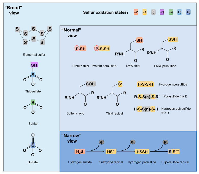
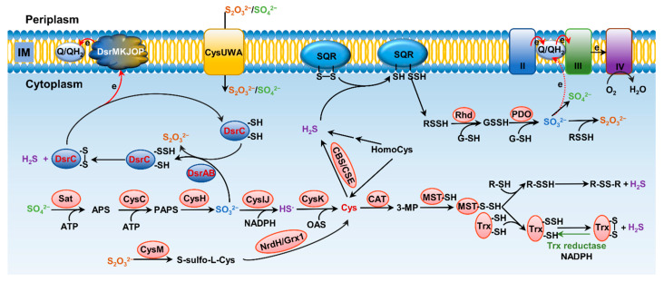
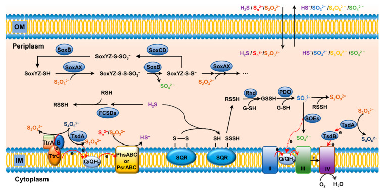
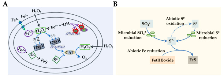
Sulfur is not only one of the most abundant elements on the Earth, but it is also essential to all living organisms. As life likely began and evolved in a hydrogen sulfide (H2S)-rich environment, sulfur metabolism represents an early form of energy generation via various reactions in prokaryotes and has driven the sulfur biogeochemical cycle since. It has long been known that H2S is toxic to cells at high concentrations, but now this gaseous molecule, at the physiological level, is recognized as a signaling molecule and a regulator of critical biological processes. Recently, many metabolites of H2S, collectively called reactive sulfur species (RSS), have been gradually appreciated as having similar or divergent regulatory roles compared with H2S in living organisms, especially mammals. In prokaryotes, even in bacteria, investigations into generation and physiology of RSS remain preliminary and an understanding of the relevant biological processes is still in its infancy. Despite this, recent and exciting advances in the fields are many. Here, we discuss abiotic and biotic generation of H2S/RSS, sulfur-transforming enzymes and their functioning mechanisms, and their physiological roles as well as the sensing and regulation of H2S/RSS.

Keywords: hydrogen sulfide; reactive sulfur species; sensing and regulation; sulfur transformation.


Conflict of interest statement

The authors declare no conflict of interest.

Figures

Figure 1
Figure 1
Structures of some RSS chemotypes in different views. The red (−2), yellow (−1), gray (0), purple (+1), green (+4), light blue (+5) and dark blue (+6) rectangles are used to designate the valence states of sulfur, as specified.
Figure 2
Figure 2
Pathways for bacterial sulfur metabolism in the cytoplasm. H2S biogenesis through amino acid metabolism: generation of H2S from homocys can be catalyzed by cystathionine β-synthase (CBS) and cystathionine γ-lyase (CSE). Cys aminotransferase (CAT) catalyzes the formation of 3-mercaptopyruvate (3-MP) from cys, and then, mercaptopyruvate sulfurtransferase (MST) converts 3-MP to H2S. H2S biogenesis occurs through assimilatory sulfate reduction (ASR) and dissimilatory sulfate reduction (DSR) of inorganic sulfur species; the latter only occurs in sulfate-reducing bacteria (SRB), catalyzing SO32− to HS through dissimilatory sulfite reductase (Dsr). For ASR pathway, SO42−, which imported from ATP-dependent transporter CysUWA, is catalyzed and converted to HS by a series of enzymes, including Sat, CysC, CysH, and CysIJ. S2O32− can also be reduced and used to synthesize cys by CysM and NrdH/Grx1. H2S catabolism: H2S binds to sulfur quinone oxidoreductase (SQR), and goes through a series of sequential reactions, including oxidation of sulfide to polysulfide by membrane-bound SQR, formation of GSSH from reaction involving with rhodanese (Rhd), oxidation of the sulfane sulfur in GSSH to SO32− catalyzed by persulfide dioxygenase (PDO), formation of S2O32− from spontaneous reaction between polysulfide and SO32−, and formation of SO42− either spontaneously or catalyzed by various enzymes, transferring two electrons via quinone into the electron transport chain.
Figure 3
Figure 3
Structure of representative enzymes involved in transformation of sulfur compounds in bacteria. (A) Overall structure of the Dsr A2B2 heterotetramer of Desulfovibrio vulgaris (PDB ID: 2V4J), green; Archaeoglobus fulgidus (PDB ID: 3MM5), yellow; Desulfovibrio gigas (PDB ID: 3OR1), pink; and Desulfoicrobrium norvegicum (PDB ID: 2XSJ), blue. Heme ligands are shown in ball-stick model. (B) Structure of TsdA of Marichromatium purpuratum (PDB ID: 5LO9; violet) and of Allochromatium vinosum (PDB ID: 4WQ7; orange). Heme ligands are shown in ball-stick model. Expanded regions show the ligands to hemes in active centers. (C) TsdA of Campylobacter jejuni (orange, prepared from AlaphaFold database [90,91]) is aligned and superimposed onto TsdAB of M. purpuratum (PDB ID: 5LO9), which are in violet and yellow, respectively. Heme ligands are shown in ball-stick model. Expanded region shows the ligands to heme 2. (D) Structure of the SoxXA (PDB ID: 2C1D) (left) and SoxYZ-B (PDB ID: 4UWQ) (right) complexes. Heme ligands are shown in ball-stick model. Expanded region shows the active site positioning at the substrate channel of SoxB.
Figure 4
Figure 4
Pathways for bacterial sulfur transformation in the periplasm. Oxidation of S2− and S0: Flavocyt c sulfide dehydrogenases (FCSDs) can oxidize H2S to the final product polysulfide. In dissimilatory sulfur-oxidizing bacteria, Rhds and PDOs are located in the periplasm, and the oxidation of H2S is catalyzed by SQRs, which consistently expose the reaction to the periplasm space. Oxidation of S0 and S2+: Unconjugated SoxYZ is catalyzed by SoxAX with S2O32−, generating SoxYZ-S-S-SO3, which is subsequently converted to SoxYZ-S-S, releasing one molecular of SO42− under the catalysis of SoxB. Reduction of S2+: TtrABC and TsdAB are responsible for interconversion of S4O62− and S2O32−. Reduction of S0: PhsABC and PsrABC are supposed to participate in the reduction of S0 (Sn2−) and S0 (S2O32−) to HS.
Figure 5
Figure 5
Physiological impacts of hydrogen sulfide. (A) H2S in oxidative stress. Upon oxidative stress, macromolecules such as DNA and proteins are damaged primarily by OH, which is generated from the interaction of Fe2+ and H2O2. H2S is a strong inhibitor of hemoproteins, catalase (CAT) in particular. With CAT inhibited, cells are unable to promptly decompose H2O2, leading to increased sensitivity to H2O2 killing. However, the prolonged presence of H2O2 activates OxyR, the master regulator in response to oxidative stress, which in turn induces expression of genes under its control, including CAT. As a result, cells gain an increased resistance against H2O2. In addition, it has been suggested that H2S from endogenous and exogenous sources may offer protection against oxidative stress by sequestering free Fe2+ intracellularly. (B) H2S in metal reduction. Microbial reduction of SO42− and elemental S0 to S2−, which catalyzes abiotic reduction of Fe3+ of Fe(III)oxide and forms FeS with the resulting Fe2+.
Figure 6
Figure 6
Structures and responding mechanism of CstR to RSS. Left, CstR (PDB ID: 7MQ2), which exists as homotetramer. Right, SqrR (PDB: 6O8N), which exists as homodimer. Expanded regions show the mechanism of activation. Both regulators use multiple cys thiols for sensing RSS by forming di-, tri-, and tetra-sulfide bonds.

Similar articles

Cited by

References

    1. Giles G.I., Tasker K.M., Jacob C. Hypothesis: The role of reactive sulfur species in oxidative stress. Free Radic. Biol. Med. 2001;31:1279–1283. doi: 10.1016/S0891-5849(01)00710-9. - DOI - PubMed
    1. Sies H., Jones D.P. Reactive oxygen species (ROS) as pleiotropic physiological signalling agents. Nat. Rev. Mol. Cell Biol. 2020;21:363–383. doi: 10.1038/s41580-020-0230-3. - DOI - PubMed
    1. Herb M., Schramm M. Functions of ROS in macrophages and antimicrobial Immunity. Antioxidants. 2021;10:313. doi: 10.3390/antiox10020313. - DOI - PMC - PubMed
    1. Forman H.J., Zhang H. Targeting oxidative stress in disease: Promise and limitations of antioxidant therapy. Nat. Rev. Drug Discov. 2021;20:689–709. doi: 10.1038/s41573-021-00233-1. - DOI - PMC - PubMed
    1. Sies H. Chapter 13—Oxidative Stress: Eustress and Distress in Redox Homeostasis. In: Fink G., editor. Stress: Physiology, Biochemistry, and Pathology. Academic Press; Cambridge, MA, USA: 2019. pp. 153–163.