

Apple Derived Exosomes Improve Collagen Type I Production and Decrease MMPs during Aging of the Skin through Downregulation of the NF-κB Pathway as Mode of Action
- PMID: 36552714
- PMCID: PMC9776931
- DOI: 10.3390/cells11243950
Apple Derived Exosomes Improve Collagen Type I Production and Decrease MMPs during Aging of the Skin through Downregulation of the NF-κB Pathway as Mode of Action
Abstract
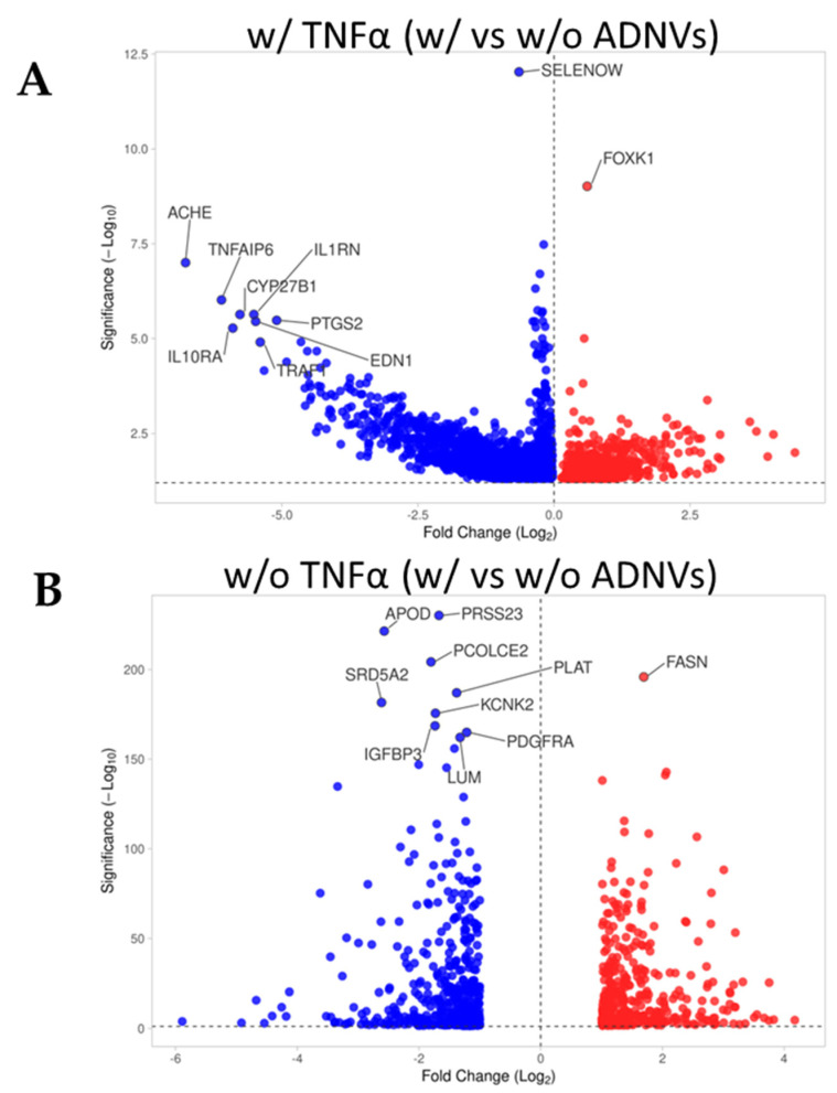
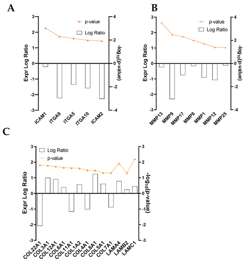
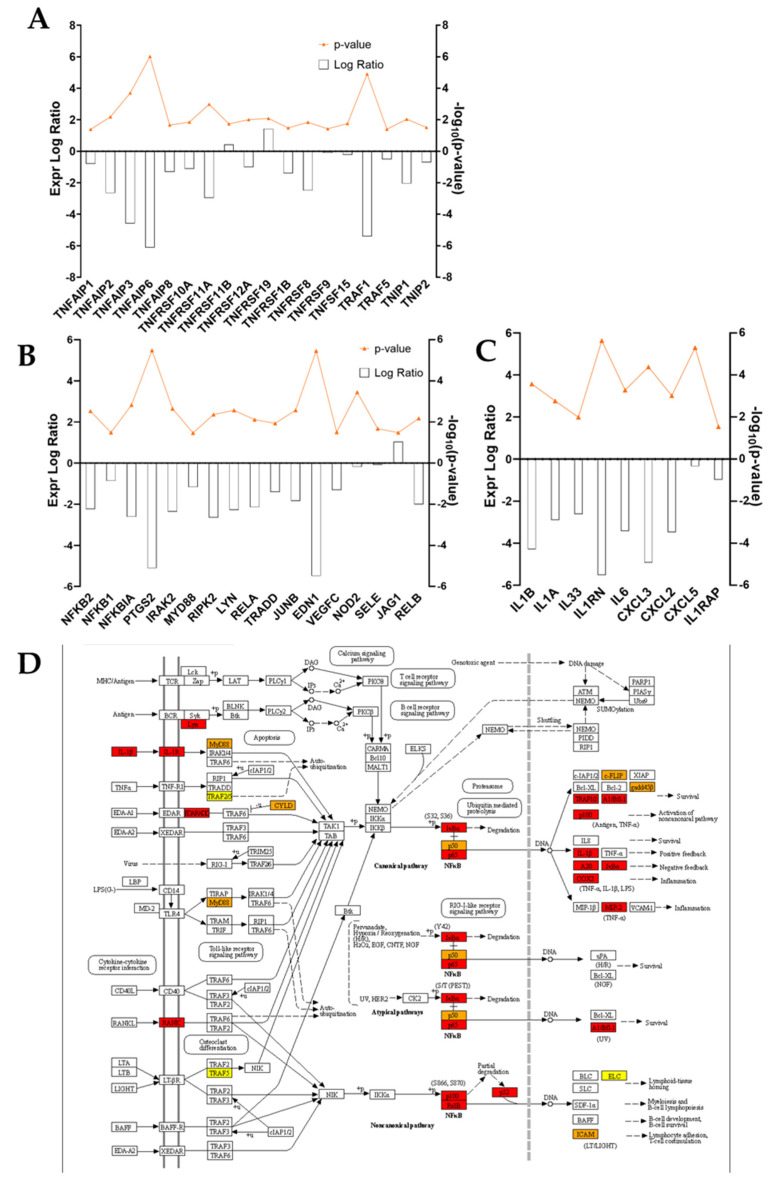
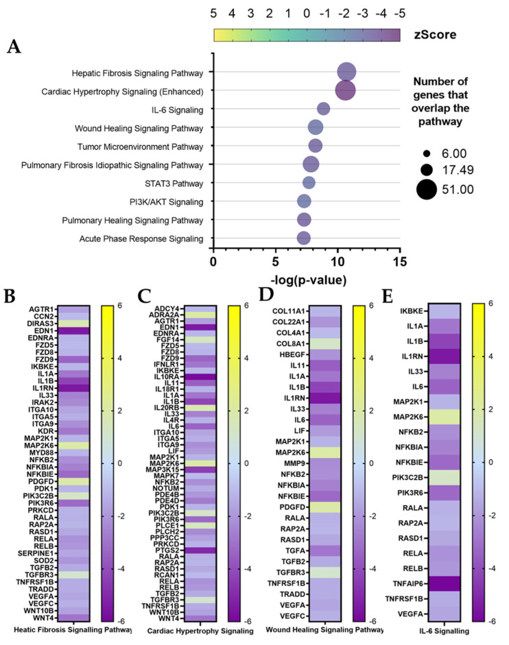
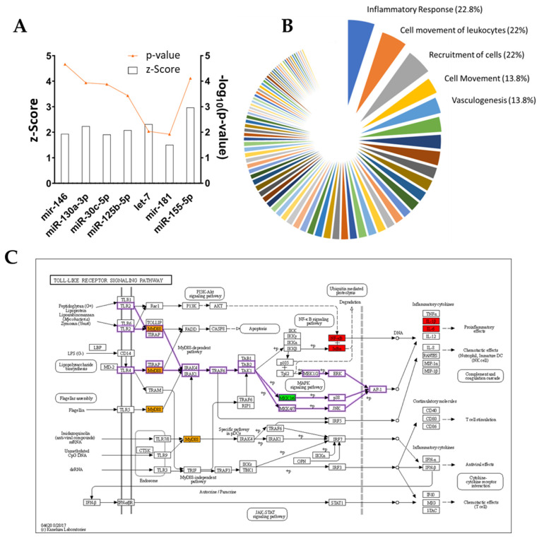
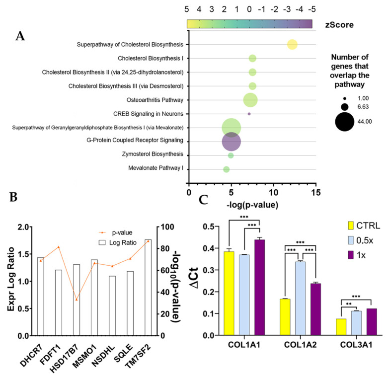
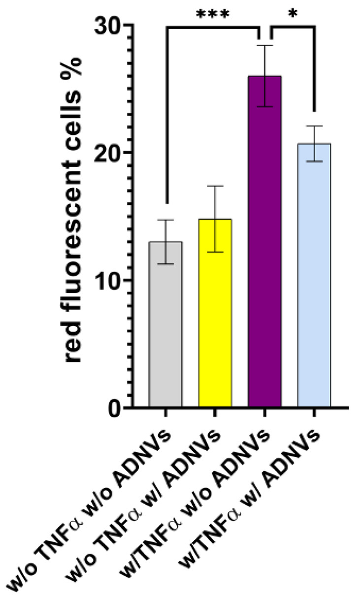
Skin ageing is strictly related to chronic inflammation of the derma and the decay of structural proteins of the extracellular matrix. Indeed, it has become common practice to refer to this phenomenon as inflammageing. Biotech innovation is always in search of new active principles that induce a youthful appearance. In this paper, apple-derived nanovesicles (ADNVs) were investigated as novel anti-inflammatory compounds, which are able to alter the extracellular matrix production of dermal fibroblasts. Total RNA sequencing analysis revealed that ADNVs negatively influence the activity of Toll-like Receptor 4 (TLR4), and, thus, downregulate the NF-κB pro-inflammatory pathway. ADNVs also reduce extracellular matrix degradation by increasing collagen synthesis (COL3A1, COL1A2, COL8A1 and COL6A1) and downregulating metalloproteinase production (MMP1, MMP8 and MMP9). Topical applications for skin regeneration were evaluated by the association of ADNVs with hyaluronic-acid-based hydrogel and patches.
Keywords: apple; exosomes; extracellular matrix; inflammageing; wound healing.
Conflict of interest statement
The authors declare no conflict of interest.
Figures
Similar articles
-
Link between organic nanovescicles from vegetable kingdom and human cell physiology: intracellular calcium signalling.J Nanobiotechnology. 2024 Feb 19;22(1):68. doi: 10.1186/s12951-024-02340-8. J Nanobiotechnology. 2024. PMID: 38369472 Free PMC article.
-
Acer tataricum subsp. ginnala Inhibits Skin Photoaging via Regulating MAPK/AP-1, NF-κB, and TGFβ/Smad Signaling in UVB-Irradiated Human Dermal Fibroblasts.Molecules. 2021 Jan 27;26(3):662. doi: 10.3390/molecules26030662. Molecules. 2021. PMID: 33513930 Free PMC article.
-
Lipoteichoic acid isolated from Lactobacillus plantarum down-regulates UV-induced MMP-1 expression and up-regulates type I procollagen through the inhibition of reactive oxygen species generation.Mol Immunol. 2015 Oct;67(2 Pt B):248-55. doi: 10.1016/j.molimm.2015.05.019. Epub 2015 Jun 6. Mol Immunol. 2015. PMID: 26059754
-
Human odontoblast culture method: the expression of collagen and matrix metalloproteinases (MMPs).Adv Dent Res. 2001 Aug;15:55-8. doi: 10.1177/08959374010150011401. Adv Dent Res. 2001. PMID: 12640741 Review.
-
Inhibition of matrix metalloproteinase expression and cellular invasion by NF-κB inhibitors of microbial origin.Biochim Biophys Acta Proteins Proteom. 2020 Jun;1868(6):140412. doi: 10.1016/j.bbapap.2020.140412. Epub 2020 Mar 14. Biochim Biophys Acta Proteins Proteom. 2020. PMID: 32179183 Review.
Cited by
-
Emerging Trends in the Application of Extracellular Vesicles as Novel Oral Delivery Vehicles for Therapeutics in Inflammatory Diseases.Int J Nanomedicine. 2024 Aug 21;19:8573-8601. doi: 10.2147/IJN.S475532. eCollection 2024. Int J Nanomedicine. 2024. PMID: 39185348 Free PMC article. Review.
-
Recent advances in dermal fibroblast senescence and skin aging: unraveling mechanisms and pioneering therapeutic strategies.Front Pharmacol. 2025 Jun 18;16:1592596. doi: 10.3389/fphar.2025.1592596. eCollection 2025. Front Pharmacol. 2025. PMID: 40606604 Free PMC article. Review.
-
Adipose-derived stem cell exosomes act as delivery vehicles of microRNAs in a dog model of chronic hepatitis.Nanotheranostics. 2024 Mar 9;8(3):298-311. doi: 10.7150/ntno.93064. eCollection 2024. Nanotheranostics. 2024. PMID: 38577321 Free PMC article.
-
Macropinocytosis and Fast Endophilin-Mediated Endocytosis Mediate Absorption of Garlic Chive-Derived Vesicle-like Nanoparticles in Human Intestinal Epithelial Cells.Mol Pharm. 2025 Jul 7;22(7):3935-3948. doi: 10.1021/acs.molpharmaceut.5c00190. Epub 2025 May 23. Mol Pharm. 2025. PMID: 40409744 Free PMC article.
-
Plant-Derived Vesicle-like Nanoparticles: The Next-Generation Drug Delivery Nanoplatforms.Pharmaceutics. 2024 Apr 26;16(5):588. doi: 10.3390/pharmaceutics16050588. Pharmaceutics. 2024. PMID: 38794248 Free PMC article. Review.
References
Publication types
MeSH terms
Substances
LinkOut - more resources
Full Text Sources
Miscellaneous
