Review
. 2022 Dec 10;11(24):3994.
doi: 10.3390/cells11243994.

PICALM and Alzheimer's Disease: An Update and Perspectives

Affiliations
Review

PICALM and Alzheimer's Disease: An Update and Perspectives

Kunie Ando et al. Cells. .

Abstract

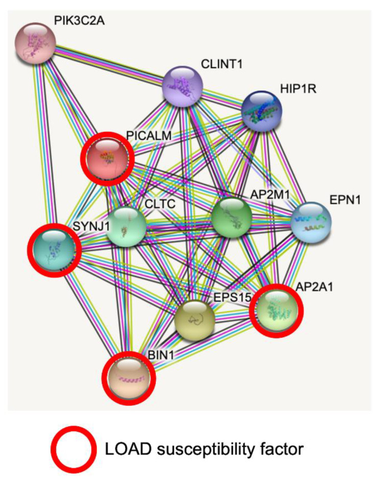
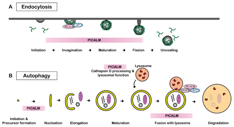
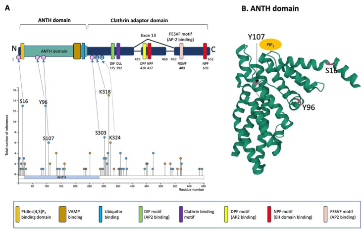
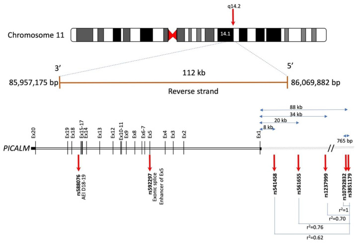
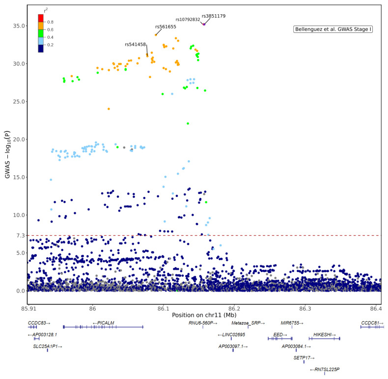
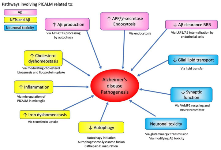
Genome-wide association studies (GWAS) have identified the PICALM (Phosphatidylinositol binding clathrin-assembly protein) gene as the most significant genetic susceptibility locus after APOE and BIN1. PICALM is a clathrin-adaptor protein that plays a critical role in clathrin-mediated endocytosis and autophagy. Since the effects of genetic variants of PICALM as AD-susceptibility loci have been confirmed by independent genetic studies in several distinct cohorts, there has been a number of in vitro and in vivo studies attempting to elucidate the underlying mechanism by which PICALM modulates AD risk. While differential modulation of APP processing and Aβ transcytosis by PICALM has been reported, significant effects of PICALM modulation of tau pathology progression have also been evidenced in Alzheimer's disease models. In this review, we summarize the current knowledge about PICALM, its physiological functions, genetic variants, post-translational modifications and relevance to AD pathogenesis.

Keywords: Alzheimer’s disease; GWAS; PICALM; amyloid β; microglia; neurofibrillary tangles.


Conflict of interest statement

The authors declare that the research was conducted in the absence of any commercial or financial relationships that could be construed as a potential conflict of interest.

Figures

Figure 1
Figure 1
PICALM interacts directly with several GWAS hits identified in LOAD. Predicted protein–protein interactions by STRING database analysis (https://string-db.org (accessed on 1 June 2022)) show several key proteins for clathrin-mediated endocytosis and intracellular trafficking as PICALM interactors [40]. AP2A1 and BIN1 have been identified as GWAS hits [41]. SNPs of SYNJ1 have significant associations with disease onset of AD [45]. CLTC: Clathrin heavy chain 1. SYNJ1: Synaptojanin1. PIK3C2A: Phosphatidylinositol 4-phosphate 3-kinase C2 domain-containing subunit α. CLINT1: Clathrin interactor 1. HIP1R: Huntingtin-interacting protein 1-related protein. AP2M1: AP2 complex subunit mu. EPN1: Epsin-1. AP2A1: AP2 complex subunit α-1. EPS15: Epidermal growth factor receptor substrate 15. BIN1: Myc box-dependent-interacting protein 1.
Figure 2
Figure 2
PICALM regulates clathrin-mediated endocytosis and autophagy. (A) PICALM is involved in clathrin-mediated endocytosis (CME) as a clathrin adaptor by facilitating the formation of clathrin-coated vesicles (CCVs). The interaction between PICALM, adaptor proteins (AP) and clathrin is critical to maintaining the form and sizes of CCVs. PICALM interacts with AP2 at the plasma membrane and AP 1 in the trans-Golgi network to form CCVs. (B) PICALM is also involved in multiple steps of autophagy process. PICALM is involved in the autophagy precursor formation at the autophagosome-lysosome fusion [51], maturation of Cathepsin D and lysosomal function [48].
Figure 3
Figure 3
Schematic illustration of PICALM structure and post-translational modifications. (A) Schematic structure of PICALM. PICALM is constituted of two distinct domains: the ANTH (AP180 N-terminal homology) domain and clathrin adaptor domain. ANTH domain contains a PtdIns(4,5)P2 binding domain and vesicle-associated membrane protein (VAMP) binding domain. Clathrin adaptor domain contains several motifs that are critical to interact with endocytic protein, including DIF, DLL Clathrin binding, DPF, NPF and FESVF motifs. DIF motif binds to AP2. DLL motif is conserved and has weak affinity to Clathrin DPF (Aspartic acid-Proline-Phenylalanine) motifs. NPF (Asparagine-Proline-Phenylalanine) motif binds to AP2 and EH domains (Eps15 Homology domain). Four phosphorylation sites (S16, Y96, S107 and S303) and two ubiquitination sites (K318 and K324) have been described in more than 5 references and validated by mass-spectrometry and thus are highlighted [65]. Altogether, the reported PTMs of PICALM are 41 phosphorylation (S2, S5, T11, S16, T18, S20, S23, T82, Y88, Y96, S107, Y138, T146, T158, Y237, S297, T301, S303, S307, S308, T312, S315, S353, T355, T356, S359, T363, T370, T379, S381, S444, S450, S453, T472, S474, K515, K534, S537, S543, T573 and S645), 15 ubiquitination (K24, K112, K134, K163, K231, K252, K288, K291, K318, K324, K336, K515, K559, K570 and K571), 1 acetylation (K28), 1 mono-methylation (R9), 1 di-methylation (R636) and 1 sumoylation (K238). (B) Computational modeling of the structure of PICALM N-terminal ANTH domain is shown (https://www.rcsb.org/structure/3ZYM (accessed on 1 June 2022)) [66]. PICALM ANTH domain is constituted of 11 α-helices. Three known phosphorylation sites (S16, Y96 and S107) in ANTH domain are highlighted in pink.
Figure 4
Figure 4
Schematic illustration of PICALM genomic location and some of the LOAD-associated SNPs based on the information available in Gene database (https://genome.ucsc.edu/ (accessed on 1 June)) and e!Ensembl (http://www.ensembl.org/ (accessed on 1 June 2022) (accession number ENSG00000073921, GRCh38:CM000673.2). Human PICALM gene spans 112 kb on chromosome 11q14 and encodes 20 exons. The most significant LOAD-associated SNP is rs3851179, located approximately 88 kb upstream of PICALM in the 5′ intergenic region [17]. rs10792832 is 765 bp from rs3851179 and in full linkage disequilibrium (LD) with rs3851179 (r2 = 1). rs1237999, located 34 kb upstream of PICALM, is localized in a CCCTC binding factor in a regulatory region [81]. rs561655 is the lead SNP of PICALM locus in [82], located 20 kb upstream of PICALM gene, a putative transcription factor binding site [15] and in LD with rs3851179 (r2 = 0.76). rs541458, located in 8 kb upstream of PICALM gene, is also genome-wide significant [15] and is in LD with rs3851179 (r2 = 0.62). rs592297 is located in exon 5 and is an exonic splice enhancer [29]. rs588076 is located in the intron 17 of PICALM and is associated with the allelic expression imbalance (AEI) in PICALM isoform lacking exons 18 and 19 (D18-19) [83].
Figure 5
Figure 5
Regional plot for PICALM locus based on Stage I results from [17] (Accession No GCST90027158, https://www.ebi.ac.uk/gwas/Study (accessed on 1 July 2022)). The plot shows the lead variant in Stage I + II meta-analysis, rs3851179 and +/− 250 kb extended coordinates for n = 3988 variants in chr11:85,907,598–86,407,598 (GRCh38). LD reference data derived from 1000 Genomes Project phased biallelic SNV and INDEL autosomal genetic variants called de novo GRCh38 (for selected n = 404 unrelated Non-Finnish European (NFE) sample) [85]. The lead variant rs3851179 is located in the intergenic region between PICALM and EED in which there are other non-coding genes (RNU6-560P, LINC02695 and Metazoa SRP) and a contigs (AP003097). rs10792832 (red) is in almost full LD with rs3851179. Other genome-wide significant SNPs, such as rs561655 [82,86] and rs541458 [15,87], are also in high LD with rs3851179. The red dashed line indicates the genome-wide significance threshold (p = 5 × 10⁻⁸).
Figure 6
Figure 6
PICALM immunolabelling is increased in LOAD and FAD in correlation with the development of tau pathology. Representative immunolabelling of anti-PICALM antibody (Sigma #HPA019053) in the Cornu Ammonis regions of the hippocampus of control (A,D), LOAD (B,E) and FAD with PSEN1 mutations at R35E and E120D (C,F). In non-demented control brain, a perinuclear immunolabelling of PICALM was detected in neurons (black arrows) (A). A PICALM immunoreactivity was detected in NFTs in neuronal perikarya (blue arrows) in the affected brain areas of LOAD (B) and FAD (C). PICALM-immunoreactive microglial cells were faintly detected in control brains (arrowheads) (D), and the number and the intensity of PICALM-stained microglia were increased in LOAD (D) (arrowheads). In FAD (F) brains, the increase in PICALM immunostained microglial cells was less prominent than in LOAD. More detailed data can be found in our article [33]. Control (Ctrl), Late-onset Alzheimer’s disease (LOAD), Familial Alzheimer’s disease (FAD). Scale bar 20 µm.
Figure 7
Figure 7
Effects of PICALM on AD pathogenesis pathways and neurodegeneration. PICALM is involved in multiple pathways of AD pathogenesis. PICALM regulates pathways related to APP processing and Aβ-related neurodegeneration (shown in pink). PICALM plays critical role related to both tau and Aβ pathologies, such as cholesterol dyshomeostasis, inflammation, iron dyshomeostasis and autophagy (yellow). PICALM is also crucial for neuronal toxicity via glial lipid transport, synaptic functions, glutaminergic transmission and modifying Aβ-toxicity (blue).

References

    1. Wong C.W., Quaranta V., Glenner G.G. Neuritic Plaques and Cerebrovascular Amyloid in Alzheimer Disease Are Antigenically Related. Proc. Natl. Acad. Sci. USA. 1985;82:8729–8732. doi: 10.1073/pnas.82.24.8729. - DOI - PMC - PubMed
    1. Brion J.P., Couck A.M., Passareiro E., Flament-Durand J. Neurofibrillary Tangles of Alzheimer’s Disease: An Immunohistochemical Study. J. Submicrosc. Cytol. 1985;17:89–96. - PubMed
    1. Duyckaerts C., Delatour B., Potier M.C. Classification and Basic Pathology of Alzheimer Disease. Acta Neuropathol. 2009;118:5–36. doi: 10.1007/s00401-009-0532-1. - DOI - PubMed
    1. Voytyuk I., De Strooper B., Chavez-Gutierrez L. Modulation of Gamma- and Beta-Secretases as Early Prevention against Alzheimer’s Disease. Biol. Psychiatry. 2018;83:320–327. doi: 10.1016/j.biopsych.2017.08.001. - DOI - PubMed
    1. Kamenetz F., Tomita T., Hsieh H., Seabrook G., Borchelt D., Iwatsubo T., Sisodia S., Malinow R. App Processing and Synaptic Function. Neuron. 2003;37:925–937. doi: 10.1016/S0896-6273(03)00124-7. - DOI - PubMed

Publication types

MeSH terms