

Novel Tuning of PMMA Orthopedic Bone Cement Using TBB Initiator: Effect of Bone Cement Extracts on Bioactivity of Osteoblasts and Osteoclasts
- PMID: 36552761
- PMCID: PMC9776632
- DOI: 10.3390/cells11243999
Novel Tuning of PMMA Orthopedic Bone Cement Using TBB Initiator: Effect of Bone Cement Extracts on Bioactivity of Osteoblasts and Osteoclasts
Abstract
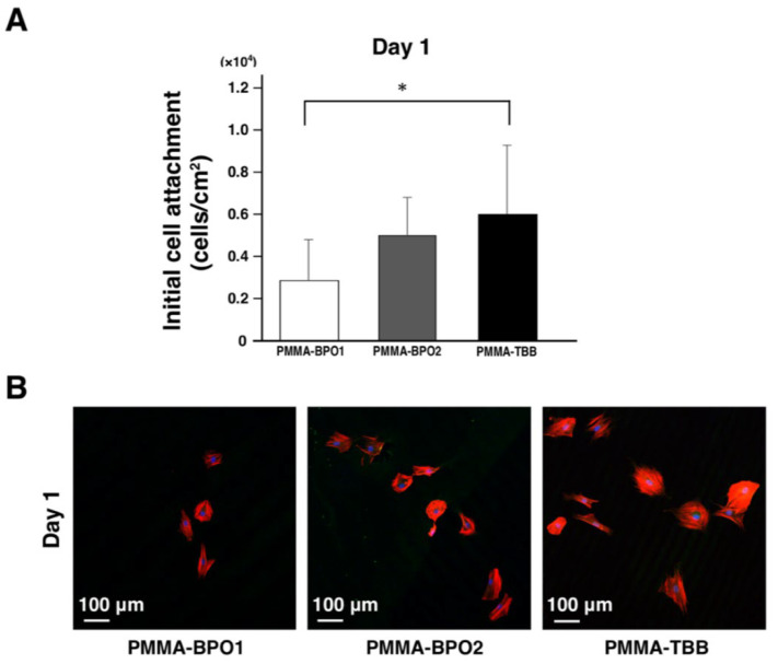
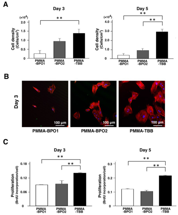
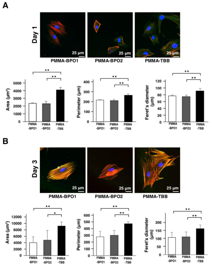
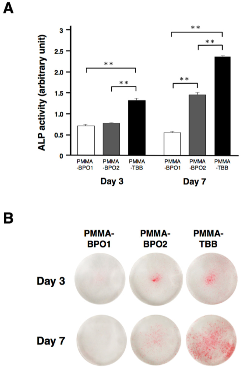
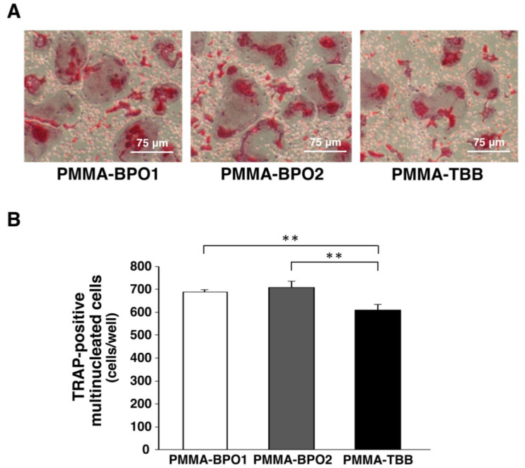
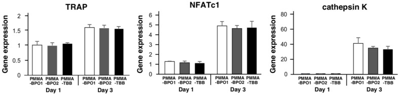
Bone cement containing benzoyl peroxide (BPO) as a polymerization initiator are commonly used to fix orthopedic metal implants. However, toxic complications caused by bone cement are a clinically significant problem. Poly (methyl methacrylate) tri-n-butylborane (PMMA-TBB), a newly developed material containing TBB as a polymerization initiator, was found to be more biocompatible than conventional PMMA-BPO bone cements due to reduced free radical generation during polymerization. However, free radicals might not be the only determinant of cytotoxicity. Here, we evaluated the response and functional phenotypes of cells exposed to extracts derived from different bone cements. Bone cement extracts were prepared from two commercial PMMA-BPO cements and an experimental PMMA-TBB. Rat bone marrow-derived osteoblasts and osteoclasts were cultured in a medium supplemented with bone cement extracts. More osteoblasts survived and attached to the culture dish with PMMA-TBB extract than in the culture with PMMA-BPO extracts. Osteoblast proliferation and differentiation were higher in the culture with PMMA-TBB extract. The number of TRAP-positive multinucleated cells was significantly lower in the culture with PMMA-TBB extract. There was no difference in osteoclast-related gene expression in response to different bone cement extracts. In conclusion, PMMA-TBB extract was less toxic to osteoblasts than PMMA-BPO extracts. Although extracts from the different cement types did not affect osteoclast function, PMMA-TBB extract seemed to reduce osteoclastogenesis, a possible further advantage of PMMA-TBB cement. These implied that the reduced radical generation during polymerization is not the only determinant for the improved biocompatibility of PMMA-TBB and that the post-polymerization chemical elution may also be important.
Keywords: arthroplasty; benzoyl peroxide; cytotoxicity; implant; poly(methyl methacrylate); total hip replacement; tri-n-butylborane.
Conflict of interest statement
The authors declare no conflict of interest.
Figures
Similar articles
-
Novel Osteogenic Behaviors around Hydrophilic and Radical-Free 4-META/MMA-TBB: Implications of an Osseointegrating Bone Cement.Int J Mol Sci. 2020 Mar 31;21(7):2405. doi: 10.3390/ijms21072405. Int J Mol Sci. 2020. PMID: 32244335 Free PMC article.
-
Prolonged Post-Polymerization Biocompatibility of Polymethylmethacrylate-Tri-n-Butylborane (PMMA-TBB) Bone Cement.Materials (Basel). 2021 Mar 8;14(5):1289. doi: 10.3390/ma14051289. Materials (Basel). 2021. PMID: 33800383 Free PMC article.
-
The Effect of TBB, as an Initiator, on the Biological Compatibility of PMMA/MMA Bone Cement.Int J Mol Sci. 2020 Jun 4;21(11):4016. doi: 10.3390/ijms21114016. Int J Mol Sci. 2020. PMID: 32512780 Free PMC article.
-
[Chemical and physical properties of PMMA bone cements].Orthopadie (Heidelb). 2023 Dec;52(12):943-956. doi: 10.1007/s00132-023-04445-8. Epub 2023 Oct 13. Orthopadie (Heidelb). 2023. PMID: 37831091 Review. German.
-
Review of the biological response to a novel bone cement containing poly(ethyl methacrylate) and n-butyl methacrylate.Biomaterials. 1998 Sep;19(17):1579-86. doi: 10.1016/s0142-9612(97)00118-x. Biomaterials. 1998. PMID: 9830983 Review.
Cited by
-
Effect of using nano-particles of magnesium oxide and titanium dioxide to enhance physical and mechanical properties of hip joint bone cement.Sci Rep. 2024 Feb 3;14(1):2838. doi: 10.1038/s41598-024-53084-2. Sci Rep. 2024. PMID: 38310142 Free PMC article.
-
Impact of Antibiotic-Loaded PMMA Spacers on the Osteogenic Potential of hMSCs.Antibiotics (Basel). 2024 Jan 3;13(1):44. doi: 10.3390/antibiotics13010044. Antibiotics (Basel). 2024. PMID: 38247603 Free PMC article.
-
Vacuum Ultraviolet (VUV) Light Photofunctionalization to Induce Human Oral Fibroblast Transmigration on Zirconia.Cells. 2023 Oct 29;12(21):2542. doi: 10.3390/cells12212542. Cells. 2023. PMID: 37947620 Free PMC article.
-
Biomaterial-based strategies for bone cement: modulating the bone microenvironment and promoting regeneration.J Nanobiotechnology. 2025 May 13;23(1):343. doi: 10.1186/s12951-025-03363-5. J Nanobiotechnology. 2025. PMID: 40361125 Free PMC article. Review.
-
Advancing osseointegration research: A dynamic three-dimensional (3D) in vitro culture model for dental implants.J Dent Sci. 2025 Jan;20(1):350-360. doi: 10.1016/j.jds.2024.06.018. Epub 2024 Jul 3. J Dent Sci. 2025. PMID: 39873044 Free PMC article.
References
-
- Gonzalez Della Valle A., Odum S.M., De A., Barrington J.W., Huddleston J.I., Illgen R.L., Springer B.D. The Effect of Femoral Fixation on Revision and Mortality Following Elective Total Hip Arthroplasty in Patients Over the Age of 65 years. An Analysis of the American Joint Replacement Registry. J. Arthroplast. 2022;37:1105–1110. doi: 10.1016/j.arth.2022.01.088. - DOI - PubMed
-
- Lewis P.L., Annette W.D., Robertsson O., Prentice H.A., Graves S.E. Impact of patient and prosthesis characteristics on common reasons for total knee replacement revision: A registry study of 36,626 revision cases from Australia, Sweden, and USA. Acta Orthop. 2022;93:623–633. doi: 10.2340/17453674.2022.3512. - DOI - PMC - PubMed
-
- Mohammad H.R., Judge A., Murray D.W. A matched comparison of the patient-reported outcome measures of cemented and cementless total knee replacements, based on the National Joint Registry of England, Wales, Northern Ireland, and Isle of Man and England’s National PROM collection programme. Acta Orthop. 2022;93:164–170. doi: 10.2340/17453674.2021.896. - DOI - PMC - PubMed
-
- Page R.S., Pai V., Eng K., Bain G., Graves S., Lorimer M. Cementless versus cemented glenoid components in conventional total shoulder joint arthroplasty: Analysis from the Australian Orthopaedic Association National Joint Replacement Registry. J. Shoulder Elb. Surg. 2018;27:1859–1865. doi: 10.1016/j.jse.2018.03.017. - DOI - PubMed
