. 2022 Dec 14;11(24):4042.
doi: 10.3390/cells11244042.

Imaging Cataract-Specific Peptides in Human Lenses

Affiliations

Imaging Cataract-Specific Peptides in Human Lenses

Kevin L Schey et al. Cells. .

Abstract

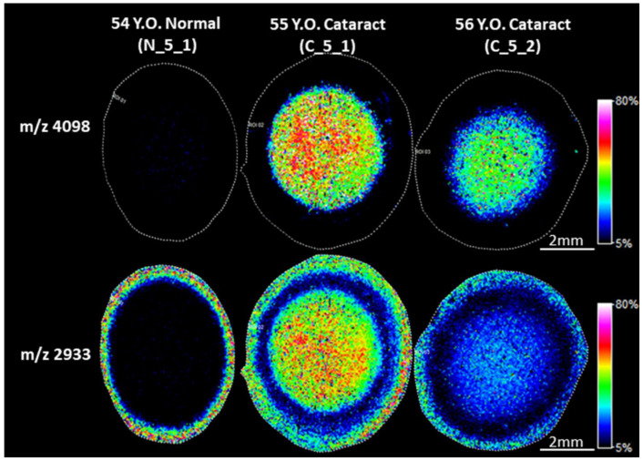
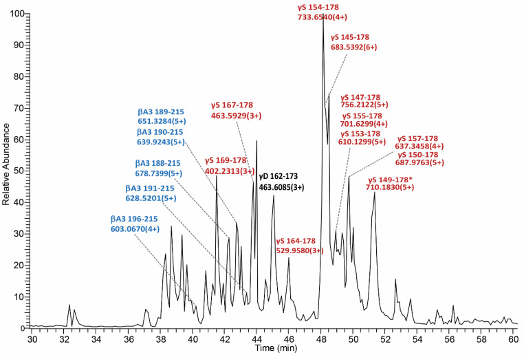
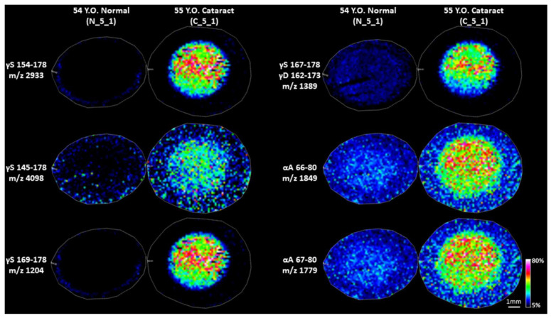
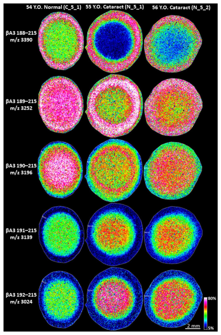
Age-related protein truncation is a common process in long-lived proteins such as proteins found in the ocular lens. Major truncation products have been reported for soluble and membrane proteins of the lens, including small peptides that can accelerate protein aggregation. However, the spatial localization of age-related protein fragments in the lens has received only limited study. Imaging mass spectrometry (IMS) is an ideal tool for examining the spatial localization of protein products in tissues. In this study we used IMS to determine the spatial localization of small crystallin fragments in aged and cataractous lenses. Consistent with previous reports, the pro-aggregatory αA-crystallin 66-80 peptide as well as αA-crystallin 67-80 and γS-crystallin 167-178 were detected in normal lenses, but found to be increased in nuclear cataract regions. In addition, a series of γS-crystallin C-terminal peptides were observed to be mainly localized to cataractous regions and barely detected in transparent lenses. Other peptides, including abundant αA3-crystallin peptides were present in both normal and cataract lenses. The functional properties of these crystallin peptides remain unstudied; however, their cataract-specific localization suggests further studies are warranted.

Keywords: cataract; imaging mass spectrometry; ocular lens; protein degradation.


Conflict of interest statement

The authors have no potential conflict of interest to declare.

Figures

Figure 1
Figure 1
Cataract-specific signals in the nucleus of cataract lenses. Imaging mass spectrometry (IMS) analysis of human lens sections was performed on Bruker Rapiflex Tissuetyper using a 60 µm raster step size. IMS analysis identified two signals with m/z values of 4098 and 2933 that were only detected in the nucleus of cataract lenses, but not in normal lens nucleus. Accurate mass measurement confirmed m/z 2933 signal in the cortex region represent a different peptide than that represented by the signal in the cataract nucleus. Signal represents peak m/z value ± 0.1% m/z unit.
Figure 2
Figure 2
Accurate mass measurement of the signal at m/z 2933 in cortex and nucleus regions using an FT-ICR mass spectrometer. The positive mode mass spectra from LCM captured cortex and nucleus samples were zoomed in to show the isotopic distribution of signals at m/z 2933. The protonated ion in a 54 Y.O. (N_5_1) normal lens cortex region (top row) has a measured monoisotopic mass of 2930.4939 Da and 2930.4999 Da in 55 Y.O. (C_5_1) cataract lens cortex (middle row), but has a measured monoisotopic mass of 2931.6001 Da in 55 Y.O. cataract lens nucleus (bottom row).
Figure 3
Figure 3
The base peak chromatogram of LC-MS/MS analysis of the extract from a 70 Y.O. (C_7_1) nucleus of a cataract lens. The base peak chromatogram is dominated by strong signals of γS- and βA3-crystallin fragments. Two strongest signals are identified as γS154–178 and γS145–178. Other base peak signals were identified as γS-crystallin C-terminal peptides (red), βA3-crystallin C-terminal peptides (blue) and a γD-crystallin C-terminal peptide (black). * indicates pyroGlu at the N-terminus.
Figure 4
Figure 4
ETD tandem mass spectra of: (A) γS-crystallin 154–178 (+4 charged ion) and (B) γS-crystallin 145–178 (+6 charged ion). All masses labeled represent the monoisotopic masses.
Figure 5
Figure 5
IMS images of LMW fragments specific or highly enriched in cataract lenses. Images were acquired on Bruker Rapiflex Tissuetyper using a 150 µm raster step size. Signal represents peak m/z value ± 0.01% m/z unit.
Figure 6
Figure 6
IMS analysis of βA3 peptides in normal and cataract lenses. IMS analysis of human lens sections was performed on Bruker Rapiflex Tissuetyper using a 60 µm raster step size. Multiple βA3-crystallin fragments were detected in normal and cataract lenses. The protein cleavage started in lens cortex and βA3 fragments were further degraded and their formation was accelerated in cataract lens nuclei. Signal represents peak m/z value ± 0.1% m/z unit.

Similar articles

Cited by

References

    1. Congdon N., Vingerling J.R., Klein B.E.K., West S., Friedman D., Kempen J., O’Colmain B., Wu S.-Y., Taylor H.R. Eye Diseases Prevalence Research Group Prevalence of Cataract and Pseudophakia/Aphakia Among Adults in the United States. Arch. Ophthalmol. 2004;122:487–494. doi: 10.1001/archopht.122.4.487. - DOI - PubMed
    1. Truscott R.J., Schey K.L., Friedrich M.G. Old Proteins in Man: A Field in its Infancy. Trends Biochem. Sci. 2016;41:654–664. doi: 10.1016/j.tibs.2016.06.004. - DOI - PMC - PubMed
    1. Takemoto L. Quantitation of C-terminal modification of alpha-A crystallin during aging of the human lens. Exp. Eye Res. 1995;60:721–724. - PubMed
    1. Wilmarth P.A., Tanner S., Dasari S., Nagalla S.R., Riviere A., Bafna V., Pevzner P.A., David L.L. Age-related changes in human crystallins determined from comparative analysis of post-translational modifications in young and aged lens: Does deamidation contribute to crystallin insolubility? J. Proteome Res. 2006;5:2554–2566. doi: 10.1021/pr050473a. - DOI - PMC - PubMed
    1. Grey A.C., Schey K.L. Age-Related Changes in the Spatial Distribution of Human Lens α-Crystallin Products by MALDI Imaging Mass Spectrometry. Investig. Opthalmology Vis. Sci. 2009;50:4319–4329. doi: 10.1167/iovs.09-3522. - DOI - PMC - PubMed

Publication types