. 2022 Dec 16;11(24):4080.
doi: 10.3390/cells11244080.

Hepatic Peroxisome Proliferator-Activated Receptor Alpha Dysfunction in Porcine Septic Shock

Affiliations

Hepatic Peroxisome Proliferator-Activated Receptor Alpha Dysfunction in Porcine Septic Shock

Jolien Vandewalle et al. Cells. .

Abstract

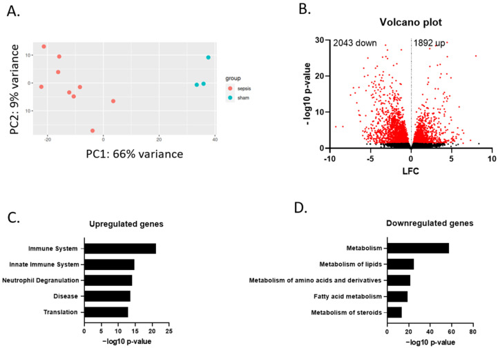
Despite decades of research, sepsis remains one of the most urgent unmet medical needs. Mechanistic investigations into sepsis have mainly focused on targeting inflammatory pathways; however, recent data indicate that sepsis should also be seen as a metabolic disease. Targeting metabolic dysregulations that take place in sepsis might uncover novel therapeutic opportunities. The role of peroxisome proliferator-activated receptor alpha (PPARɑ) in liver dysfunction during sepsis has recently been described, and restoring PPARɑ signaling has proven to be successful in mouse polymicrobial sepsis. To confirm that such therapy might be translated to septic patients, we analyzed metabolic perturbations in the liver of a porcine fecal peritonitis model. Resuscitation with fluids, vasopressor, antimicrobial therapy and abdominal lavage were applied to the pigs in order to mimic human clinical care. By using RNA-seq, we detected downregulated PPARɑ signaling in the livers of septic pigs and that reduced PPARɑ levels correlated well with disease severity. As PPARɑ regulates the expression of many genes involved in fatty acid oxidation, the reduced expression of these target genes, concomitant with increased free fatty acids in plasma and ectopic lipid deposition in the liver, was observed. The results obtained with pigs are in agreement with earlier observations seen in mice and support the potential of targeting defective PPARɑ signaling in clinical research.

Keywords: PPARɑ; free fatty acids; metabolism; sepsis; swine.


Conflict of interest statement

The authors declare no conflict of interest.

Figures

Figure 1
Figure 1
Protocol timeline. Animals were allowed to develop sepsis until a severe hypotension with mean arterial pressure (MAP) ≤ 50 mmHg was obtained. Severe hypotension (MAP between 45 and 50 mmHg) was allowed for one hour. Thereafter, full resuscitation was started, aiming to restore the pulse pressure variation (PPV) to ≤13% or MAP ≥ 65 mmHg. After achieving this objective, antibiotics and abdominal lavage were applied to control the infection. Norepinephrine was added to the infusion to maintain a MAP of 65–75 mmHg for 8 h. After this timepoint, the animals were euthanized to collect liver and plasma for subsequent analysis (i.e., ~18 h after the start of the experiment).
Figure 2
Figure 2
Metabolic dysregulation in livers of porcine sepsis. (A) PCA plot of overall gene expression in liver samples from sham and sepsis pigs. PCA plot clarifies the variance within samples per condition. (B) Volcano plot with differential expression of genes affected by sepsis compared to sham, as measured by bulk RNA sequencing. Genes significantly affected are indicated with red dots (p < 0.05). Genes with −log10 p-value above 30 are omitted for clarity of the plot. (C) Top five enriched gene ontology (GO) terms for genes that are upregulated upon sepsis (p < 0.05). (D) Top five enriched GO terms for genes that are downregulated upon sepsis (p < 0.05). Analyses of (C,D) was performed with the Enrichr tool.
Figure 3
Figure 3
PPARα dysfunction in livers of porcine sepsis. (A) Top five enriched gene ontology (GO) terms for genes that are downregulated upon sepsis (p < 0.05). Analysis was performed with the Wiki pathway tool. (B) Homer motif analysis to detect DNA binding motifs in the 500 base pairs upstream of the transcription start site of up- and down regulated genes upon sepsis (p < 0.05 and for genes with a mouse orthologue). Top five motifs ranked according to p-value is shown. (C) Violin plot showing LFC upon sepsis of genes that are known to be induced by the PPARα agonist GW7647 in murine sepsis (p < 0.05). LFC of the porcine genes with a mouse orthologue upon sepsis is displayed (389 genes). Median and upper and lower quartiles are shown with a horizontal dotted line in the violin plot. Median is significantly different from baseline LFC 0. **** p < 0.0001. (D) Violin plot showing LFC upon sepsis of genes involved in FA oxidation (GO00193951, 121 genes) that are known to be induced by the PPARα agonist GW7647 (34/121) in murine sepsis (p < 0.05). LFC of the porcine genes with a mouse orthologue upon sepsis is displayed (29 genes). Median and upper and lower quartiles are shown with a horizontal dotted line in the violin plot. Median is significantly different from baseline LFC 0. **** p < 0.0001. (E) Expression level of PPARα and genes involved in β-oxidation of FFAs and ketogenesis is displayed. The expression level of sham pigs is set as 100% and compared to the level in sepsis pigs. (F) Correlation curve plotting the norepinephrine (NE) requirement of each septic pig to their PPARα mRNA level in the liver (RNA-seq counts are log2 normalized). Data is analyzed with a simple linear regression. R2 depicts correlation of 0.6955. The slope is significantly different from zero. N = 9.
Figure 4
Figure 4
Metabolic disease parameters in blood and liver of porcine sepsis. (A) Concentration FFA in plasma of sham (n = 3) and sepsis (n = 9) pigs. (B) Immunofluorescent images of porcine liver after sham or sepsis. Cryosections were stained with Hoechst (blue), acti-stain (green) and lipidTOX (red). Z-stacks (8/region) were generated in 3 areas scattered across the entire tissue section. White scale bar = 50 μm. One representative picture of each biological repeat is shown. (C) The amount of lipid droplets (LDs)/cell were calculated for each Z-stack. Averages of the amount of the LDs were converged for each subject and biological replicates (n = 2) are used in the figure.

References

    1. Singer M., Deutschman C.S., Seymour C., Shankar-Har M., Annane D., Angus D.C. The Third International Consensus Definitions for Sepsis and Septic Shock (Sepsis-3) JAMA. 2016;315:801–810. doi: 10.1001/jama.2016.0287. - DOI - PMC - PubMed
    1. Rudd K.E., Johnson S.C., Agesa K.M., Shackelford K.A., Tsoi D., Kievlan D.R., Colombara D.v., Ikuta K.S., Kissoon N., Finfer S., et al. Global, Regional, and National Sepsis Incidence and Mortality, 1990–2017: Analysis for the Global Burden of Disease Study. Lancet. 2020;395:200–211. doi: 10.1016/S0140-6736(19)32989-7. - DOI - PMC - PubMed
    1. Cavaillon J., Singer M., Skirecki T. Sepsis Therapies: Learning from 30 Years of Failure of Translational Research to Propose New Leads. EMBO Mol. Med. 2020;12:e10128. doi: 10.15252/emmm.201810128. - DOI - PMC - PubMed
    1. van Wyngene L., Vandewalle J., Libert C. Reprogramming of Basic Metabolic Pathways in Microbial Sepsis: Therapeutic Targets at Last? EMBO Mol. Med. 2018;10:e8712. doi: 10.15252/emmm.201708712. - DOI - PMC - PubMed
    1. Evans L., Rhodes A., Alhazzani W., Antonelli M., Coopersmith C.M., French C., Machado F.R., Mcintyre L., Ostermann M., Prescott H.C., et al. Surviving Sepsis Campaign: International Guidelines for Management of Sepsis and Septic Shock 2021. Intensive Care Med. 2021;47:1181–1247. doi: 10.1007/s00134-021-06506-y. - DOI - PMC - PubMed

Publication types