Investigation of Neutrophil Extracellular Traps as Potential Mediators in the Pathogenesis of Non-Acute Subdural Hematomas: A Pilot Study
- PMID: 36552941
- PMCID: PMC9776444
- DOI: 10.3390/diagnostics12122934
Investigation of Neutrophil Extracellular Traps as Potential Mediators in the Pathogenesis of Non-Acute Subdural Hematomas: A Pilot Study
Abstract
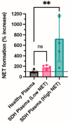
Non-acute subdural hematomas (NASHs) are a cause of significant morbidity and mortality, particularly with recurrences. Although recurrence is believed to involve a disordered neuroinflammatory cascade involving vascular endothelial growth factor (VEGF), this pathway has yet to be completely elucidated. Neutrophil extracellular traps (NETs) are key factors that promote inflammation/apoptosis and can be induced by VEGF. We investigated whether NETs are present in NASH membranes, quantified NET concentrations, and examined whether NET and VEGF levels are correlated in NASHs. Samples from patients undergoing NASH evacuation were collected during surgery and postoperatively at 24 and 48 h. Fluid samples and NASH membranes were analyzed for levels of VEGF, NETs, and platelet activation. NASH samples contained numerous neutrophils positive for NET formation. Myeloperoxidase-DNA complexes (a marker of NETs) remained elevated 48 h postoperatively (1.06 ± 0.22 day 0, 0.72 ± 0.23 day 1, and 0.83 ± 0.33 day 2). VEGF was also elevated in NASHs (7.08 ± 0.98 ng/mL day 0, 3.40 ± 0.68 ng/mL day 1, and 6.05 ± 1.8 ng/mL day 2). VEGF levels were significantly correlated with myeloperoxidase-DNA levels. These results show that NETs are increasing in NASH, a finding that was previously unknown. The strong correlation between NET and VEGF levels indicates that VEGF may be an important mediator of NET-related inflammation in NASH.
Keywords: VEGF; neutrophil extracellular trap; recurrence; subdural hematoma; vascular endothelial growth factor.
Conflict of interest statement
The authors declare no conflict of interest.
Figures


Similar articles
-
Neutrophil Extracellular Traps Promote Angiogenesis: Evidence From Vascular Pathology in Pulmonary Hypertension.Arterioscler Thromb Vasc Biol. 2016 Oct;36(10):2078-87. doi: 10.1161/ATVBAHA.116.307634. Epub 2016 Jul 28. Arterioscler Thromb Vasc Biol. 2016. PMID: 27470511
-
Neutrophil extracellular traps (NETs) are increased in the alveolar spaces of patients with ventilator-associated pneumonia.Crit Care. 2018 Dec 27;22(1):358. doi: 10.1186/s13054-018-2290-8. Crit Care. 2018. PMID: 30587204 Free PMC article.
-
Neutrophil Extracellular Traps in Asthma: Friends or Foes?Cells. 2022 Nov 7;11(21):3521. doi: 10.3390/cells11213521. Cells. 2022. PMID: 36359917 Free PMC article. Review.
-
Middle meningeal artery embolization treatment of nonacute subdural hematomas in the elderly: a multiinstitutional experience of 151 cases.Neurosurg Focus. 2020 Oct;49(4):E5. doi: 10.3171/2020.7.FOCUS20518. Neurosurg Focus. 2020. PMID: 33002874
-
Neutrophil Extracellular Traps in ANCA-Associated Vasculitis.Front Immunol. 2016 Jun 30;7:256. doi: 10.3389/fimmu.2016.00256. eCollection 2016. Front Immunol. 2016. PMID: 27446086 Free PMC article. Review.
References
-
- Hara M., Tamaki M., Aoyagi M., Ohno K. Possible role of cyclooxygenase-2 in developing chronic subdural hematoma. J. Med. Dent. Sci. 2009;56:101–106. - PubMed
Grants and funding
LinkOut - more resources
Full Text Sources
Research Materials

