

Left Ventricle Wall Motion Analysis with Real-Time MRI Feature Tracking in Heart Failure Patients: A Pilot Study
- PMID: 36552955
- PMCID: PMC9776889
- DOI: 10.3390/diagnostics12122946
Left Ventricle Wall Motion Analysis with Real-Time MRI Feature Tracking in Heart Failure Patients: A Pilot Study
Abstract
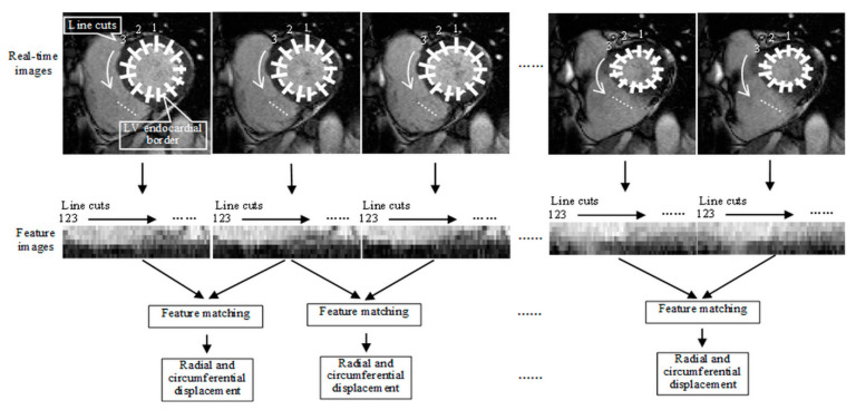
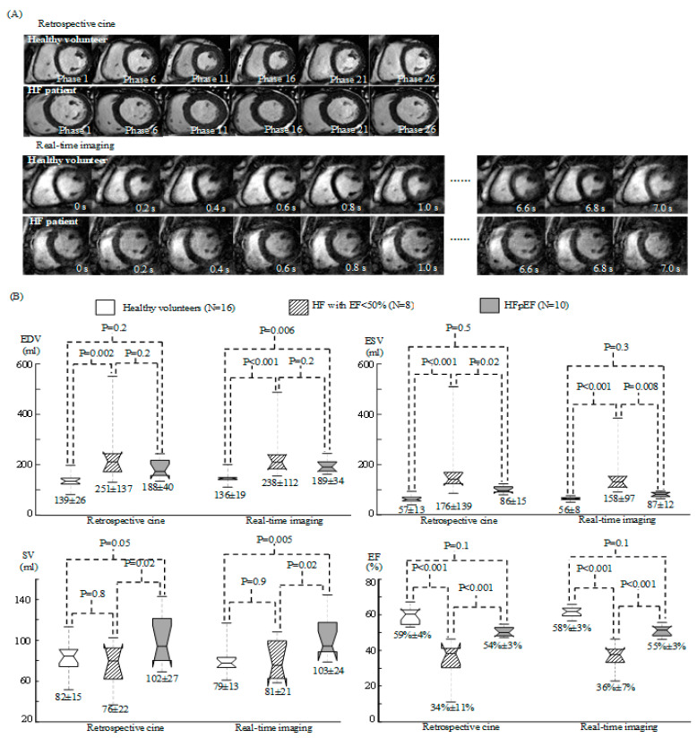
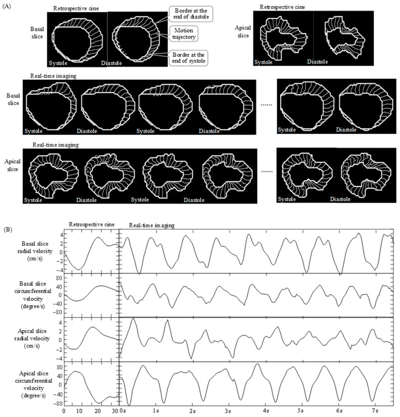
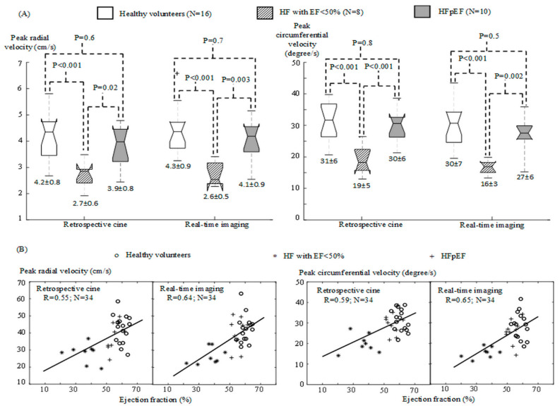
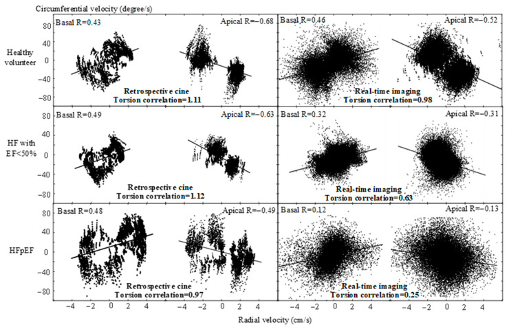
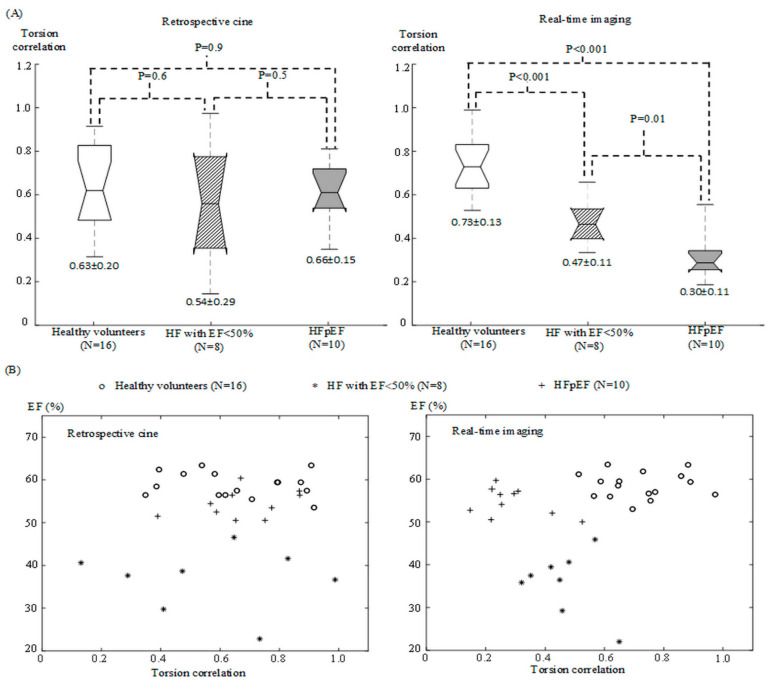
Volumetric measurements with cardiac magnetic resonance imaging (MRI) are effective for evaluating heart failure (HF) with systolic dysfunction that typically induces a lower ejection fraction (EF) than normal (<50%) while they are not sensitive to diastolic dysfunction in HF patients with preserved EF (≥50%). This work is to investigate whether HF evaluation with cardiac MRI can be improved with real-time MRI feature tracking. In a cardiac MRI study, we recruited 16 healthy volunteers, 8 HF patients with EF < 50% and 10 HF patients with preserved EF. Using real-time feature tracking, a cardiac MRI index, torsion correlation, was calculated which evaluated the correlation of torsional and radial wall motion in the left ventricle (LV) over a series of sequential cardiac cycles. The HF patients with preserved EF and the healthy volunteers presented significant difference in torsion correlation (one-way ANOVA, p < 0.001). In the scatter plots of EF against torsion correlation, the HF patients with EF < 50%, the HF patients with preserved EF and the healthy volunteers were well differentiated, indicating that real-time MRI feature tracking provided LV function assessment complementary to volumetric measurements. This study demonstrated the potential of cardiac MRI for evaluating both systolic and diastolic dysfunction in HF patients.
Keywords: LV wall motion; feature tracking; heart failure; real-time imaging; volumetric measurements.
Conflict of interest statement
The authors declare no conflict of interest.
Figures
Similar articles
-
Association Between Heart Failure With Preserved Left Ventricular Ejection Fraction and Impaired Left Atrial Phasic Function in Hypertrophic Cardiomyopathy: Evaluation by Cardiac MRI Feature Tracking.J Magn Reson Imaging. 2022 Jul;56(1):248-259. doi: 10.1002/jmri.28000. Epub 2021 Nov 19. J Magn Reson Imaging. 2022. PMID: 34799953
-
Optical Flow Analysis of Left Ventricle Wall Motion with Real-Time Cardiac Magnetic Resonance Imaging in Healthy Subjects and Heart Failure Patients.Ann Biomed Eng. 2022 Feb;50(2):195-210. doi: 10.1007/s10439-022-02907-2. Epub 2022 Jan 12. Ann Biomed Eng. 2022. PMID: 35022866
-
Temporospatial characterization of ventricular wall motion with real-time cardiac magnetic resonance imaging in health and disease.Sci Rep. 2022 Mar 8;12(1):4070. doi: 10.1038/s41598-022-08094-3. Sci Rep. 2022. PMID: 35260729 Free PMC article.
-
Contribution of systolic and diastolic abnormalities to heart failure with a normal and a reduced ejection fraction.Prog Cardiovasc Dis. 2007 Jan-Feb;49(4):229-40. doi: 10.1016/j.pcad.2006.08.009. Prog Cardiovasc Dis. 2007. PMID: 17185111 Review.
-
Heart failure diagnosis: the role of echocardiography and magnetic resonance imaging.Front Biosci (Landmark Ed). 2009 Jan 1;14(7):2688-703. doi: 10.2741/3406. Front Biosci (Landmark Ed). 2009. PMID: 19273228 Review.
References
-
- Yancy C.W., Jessup M., Bozkurt B., Butler J., Casey D.E., Jr., Colvin M.M., Drazner M.H., Filippatos G.S., Fonarow G.C., Givertz M.M., et al. 2017 ACC/AHA/HFSA Focused Update of the 2013 ACCF/AHA Guideline for the Management of Heart Failure: A Report of the American College of Cardiology/American Heart Association Task Force on Clinical Practice Guidelines and the Heart Failure Society of America. Circulation. 2017;136:e137–e161. doi: 10.1161/CIR.0000000000000509. - DOI - PubMed
-
- Ponikowski P., Voors A.A., Anker S.D., Bueno H., Cleland J.G.F., Coats A.J.S., Falk V., Gonzalez-Juanatey J.R., Harjola V.P., Jankowska E.A., et al. 2016 ESC Guidelines for the diagnosis and treatment of acute and chronic heart failure: The Task Force for the diagnosis and treatment of acute and chronic heart failure of the European Society of Cardiology (ESC)Developed with the special contribution of the Heart Failure Association (HFA) of the ESC. Eur. Heart J. 2016;37:2129–2200. doi: 10.1093/eurheartj/ehw128. - DOI - PubMed
-
- King M., Kingery J.E., Casey B. Diagnosis and evaluation of heart failure. Am. Fam. Physician. 2012;85:1161–1168. - PubMed
-
- Schulz-Menger J., Bluemke D.A., Bremerich J., Flamm S.D., Fogel M.A., Friedrich M.G., Kim R.J., von Knobelsdorff-Brenkenhoff F., Kramer C.M., Pennell D.J., et al. Standardized image interpretation and post-processing in cardiovascular magnetic resonance-2020 update: Society for Cardiovascular Magnetic Resonance (SCMR): Board of Trustees Task Force on Standardized Post-Processing. J. Cardiovasc. Magn. Reson. 2020;22:19. doi: 10.1186/s12968-020-00610-6. - DOI - PMC - PubMed
LinkOut - more resources
Full Text Sources
Research Materials
Miscellaneous
