Review
. 2022 Nov 26;12(12):2960.
doi: 10.3390/diagnostics12122960.

Advances in Imaging for Assessing Pelvic Endometriosis

Affiliations
Review

Advances in Imaging for Assessing Pelvic Endometriosis

Stefano Guerriero et al. Diagnostics (Basel). .

Abstract

In recent years, due to the development of standardized diagnostic protocols associated with an improvement in the associated technology, the diagnosis of pelvic endometriosis using imaging is becoming a reality. In particular, transvaginal ultrasound and magnetic resonance are today the two imaging techniques that can accurately identify the majority of the phenotypes of endometriosis. This review focuses not only on these most common imaging modalities but also on some additional radiological techniques that were proposed for rectosigmoid colon endometriosis, such as double-contrast barium enema, rectal endoscopic ultrasonography, multidetector computed tomography enema, computed tomography colonography and positron emission tomography-computed tomography with 16α-[18F]fluoro-17β-estradiol.

Keywords: endometriosis; magnetic resonance imaging; transvaginal ultrasound.


Conflict of interest statement

All authors report no conflict of interest/disclosures.

Figures

Figure 1
Figure 1
TVUS imaging method: a bladder endometriotic nodule with 2D TVUS (black and white images) and with 3D TVUS (color image).
Figure 2
Figure 2
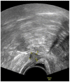
TVUS imaging method: the arrow and calipers show a nodule of the rectovaginal space in the sagittal plane.
Figure 3
Figure 3
TVUS imaging method: USL lesion indicated by an arrow and calipers in the sagittal plane. U: uterus.
Figure 4
Figure 4
TVUS imaging method: the arrow and the calipers show a posterior vaginal fornix nodule in the sagittal plane. U: uterus.
Figure 5
Figure 5
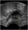
TVUS imaging method: arrow 1 shows a rectosigmoid nodule, while arrow 2 shows a USL nodule in the sagittal plane.
Figure 6
Figure 6
MRI imaging method: an endometrioma indicated by an arrow in the axial plane.
Figure 7
Figure 7
MRI imaging method: a retrocervical nodule indicated by an arrow in the sagittal plane.
Figure 8
Figure 8
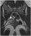
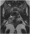
MRI imaging method: a nodule of rectosigmoid colon between the two arrows in the coronal plane.
Figure 9
Figure 9
MRI imaging method: a nodule of the USL between the two arrows in the coronal plane.

Similar articles

Cited by

References

    1. Taylor H.S., Kotlyar A.M., A Flores V. Endometriosis is a chronic systemic disease: Clinical challenges and novel innovations. Lancet. 2021;397:839–852. doi: 10.1016/S0140-6736(21)00389-5. - DOI - PubMed
    1. Tomassetti C., Johnson N.P., Petrozza J. An International Terminology for Endometriosis, 2021. J. Minim. Invasive Gynecol. 2021;28:1849–1859. doi: 10.1016/j.jmig.2021.08.032. - DOI - PubMed
    1. Exacoustos C., De Felice G., Pizzo A., Morosetti G., Lazzeri L., Centini G., Piccione E., Zupi E. Isolated Ovarian Endometrioma: A History Between Myth and Reality. J. Minim. Invasive Gynecol. 2018;25:884–891. doi: 10.1016/j.jmig.2017.12.026. - DOI - PubMed
    1. Menni K., Facchetti L., Cabassa P. Extragenital endometriosis: Assessment with MR imaging. A pictorial review. Br. J. Radiol. 2016;89:20150672. doi: 10.1259/bjr.20150672. - DOI - PMC - PubMed
    1. Andres M.P., Arcoverde F., Souza C.C., Fernandes L.F.C., Abrão M.S., Kho R.M. Extrapelvic Endometriosis: A Systematic Review. J. Minim. Invasive Gynecol. 2019;27:373–389. doi: 10.1016/j.jmig.2019.10.004. - DOI - PubMed