. 2022 Nov 30;12(12):2995.
doi: 10.3390/diagnostics12122995.

Predicting IDH Mutation Status in Low-Grade Gliomas Based on Optimal Radiomic Features Combined with Multi-Sequence Magnetic Resonance Imaging

Affiliations

Predicting IDH Mutation Status in Low-Grade Gliomas Based on Optimal Radiomic Features Combined with Multi-Sequence Magnetic Resonance Imaging

Ailing He et al. Diagnostics (Basel). .

Abstract

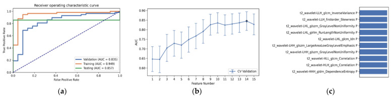
The IDH somatic mutation status is an important basis for the diagnosis and classification of gliomas. We proposed a "6-Step" general radiomics model to noninvasively predict the IDH mutation status by simultaneously tuning combined multi-sequence MRI and optimizing the full radiomics processing pipeline. Radiomic features (n = 3776) were extracted from multi-sequence MRI (T1, T2, FLAIR, and T1Gd) in low-grade gliomas (LGGs), and a total of 45,360 radiomics pipeline were investigated according to different settings. The predictive ability of the general radiomics model was evaluated with regards to accuracy, stability, and efficiency. Based on numerous experiments, we finally reached an optimal pipeline for classifying IDH mutation status, namely the T2+FLAIR combined multi-sequence with the wavelet image filter, mean data normalization, PCC dimension reduction, RFE feature selection, and SVM classifier. The mean and standard deviation of AUC, accuracy, sensitivity, and specificity were 0.873 ± 0.05, 0.876 ± 0.09, 0.875 ± 0.11, and 0.877 ± 0.15, respectively. Furthermore, 14 radiomic features that best distinguished the IDH mutation status of the T2+FLAIR multi-sequence were analyzed, and the gray level co-occurrence matrix (GLCM) features were shown to be of high importance. Apart from the promising prediction of the molecular subtypes, this study also provided a general tool for radiomics investigation.

Keywords: IDH; glioma; machine learning; multi-sequence MRI; radiomics.


Conflict of interest statement

The authors declare no conflict of interest.

Figures

Figure 1
Figure 1
Patient screening flowchart. Abbreviations: TCGA, The Cancer Genome Atlas; T1, T1-weighted; T2, T2-weighted; FLAIR, fluid-attenuated inversion recovery; and T1Gd, post-contrast T1-weighted.
Figure 2
Figure 2
Flow chart of the “6-Step” general radiomics model. The data frames in blue indicate that we chose one of the displayed methods, and the data frames in green mean that we chose all of the displayed methods. A, B, C, and D indicate different medical images. The “+” symbol indicates that different MRI sequences were combined to form a new input object. Abbreviations: GLCM, Gray-level co-occurrence matrix; GLSZM, Gray-level size zone matrix; GLRLM, Gray-level run length matrix; GLDM, Gray-level dependence matrix; and NGTDM, neighboring gray tone difference matrix.
Figure 3
Figure 3
The best performance generated by the “6-Step” general radiomics model was the T2+FLAIR combined multi-sequence with the following settings: wavelet image filter, mean data normalization, PCC dimension reduction, RFE feature selection, and SVM classifier. (a) Receiver operating characteristic (ROC) curves of the training, testing, and validation sets; (b) the FAE software’s suggestion of a candidate 14-feature model according to the “one-standard error” rule; (c) the 14 radiomic features with the highest average feature importance calculated by the best settings with the T2+FLAIR combined multi-sequence.
Figure 4
Figure 4
The best diagnostic performance of a single radiomic sequence was T2 with the following settings: wavelet image filter, mean data normalization, PCC dimension reduction, RFE feature selection, and LDA classifier. (a) Receiver operating characteristic (ROC) curves of the training, testing, and validation sets; (b) FAE software’s suggestion of a candidate 10-feature model according to the “one-standard error” rule; (c) the 10 radiomic features with the highest average feature importance calculated by the best settings with the T2 sequence.
Figure 5
Figure 5
The best diagnostic performance of the three combined multi-sequence was the T1+T2+T1Gd combined multi-sequence with the following settings: wavelet image filter, Z-score data normalization, PCA dimension reduction, RFE feature selection, and RF classifier. (a) Receiver operating characteristic (ROC) curves of the training, testing, and validation sets; (b) FAE software’s suggestion of a candidate 11-feature model according to the “one-standard error” rule; (c) the 11 radiomic features with the highest average feature importance calculated by the best settings with the T1+T2+T1Gd combined multi-sequence.
Figure 6
Figure 6
The best performance generated by the T1+T2+FLAIR+T1Gd combined multi-sequence with the following settings: wavelet image filter, mean data normalization, PCC dimension reduction, ANOVA feature selection, and RF classifier. (a) Receiver operating characteristic (ROC) curves of the training, testing and validation sets; (b) FAE software’s suggestion of a candidate 5-feature model according to the “one-standard error” rule; (c) the 5 radiomic features with the highest average feature importance calculated by the best settings with the T1+T2+FLAIR+T1Gd combined multi-sequence.
Figure 7
Figure 7
The performance with a different number of features, ranging from 1 to 15. This result was generated by the following radiomic pipeline settings: wavelet image filter, mean data normalization, PCC dimension reduction, RFE feature selection, and the SVM classifier with a number of features ranging from 1 to 15. Abbreviations: AUC, area under the curve; Acc, accuracy; Sen, sensitivity; and Spe, specificity.

References

    1. Yang K., Wu Z., Zhang H., Zhang N., Wu W., Wang Z., Dai Z., Zhang X., Zhang L., Peng Y., et al. Glioma targeted therapy: Insight into future of molecular approaches. Mol. Cancer. 2022;21:39. doi: 10.1186/s12943-022-01513-z. - DOI - PMC - PubMed
    1. Choi Y.S., Ahn S.S., Chang J.H., Kang S.-G., Kim E.H., Kim S.H., Jain R., Lee S.-K. Machine learning and radiomic phenotyping of lower grade gliomas: Improving survival prediction. Eur. Radiol. 2020;307:3834–3842. doi: 10.1007/s00330-020-06737-5. - DOI - PubMed
    1. Galbraith K., Snuderl M. Molecular Pathology of Gliomas. Surg. Pathol. Clin. 2021;14:379–386. doi: 10.1016/j.path.2021.05.003. - DOI - PubMed
    1. Weller M., van den Bent M., Tonn J.C. European Association for Neuro-Oncology (EANO) guideline on the diagnosis and treatment of adult astrocytic and oligodendroglial gliomas. Lancet Oncol. 2017;18:e315–e329. doi: 10.1016/S1470-2045(17)30194-8. - DOI - PubMed
    1. Wang L.-B., Karpova A., Gritsenko M.A., Kyle J.E., Cao S., Li Y., Rykunov D., Colaprico A., Rothstein J.H., Hong R., et al. Clinical Proteomic Tumor Analysis Consortium. Proteogenomic and metabolomic characterization of human glioblastoma. Cancer Cell. 2021;39:509–528. doi: 10.1016/j.ccell.2021.01.006. - DOI - PMC - PubMed

LinkOut - more resources