

Diffusion Tensor and Dynamic Contrast-Enhanced Magnetic Resonance Imaging Correlate with Molecular Markers of Inflammation in the Synovium
- PMID: 36553048
- PMCID: PMC9776499
- DOI: 10.3390/diagnostics12123041
Diffusion Tensor and Dynamic Contrast-Enhanced Magnetic Resonance Imaging Correlate with Molecular Markers of Inflammation in the Synovium
Abstract
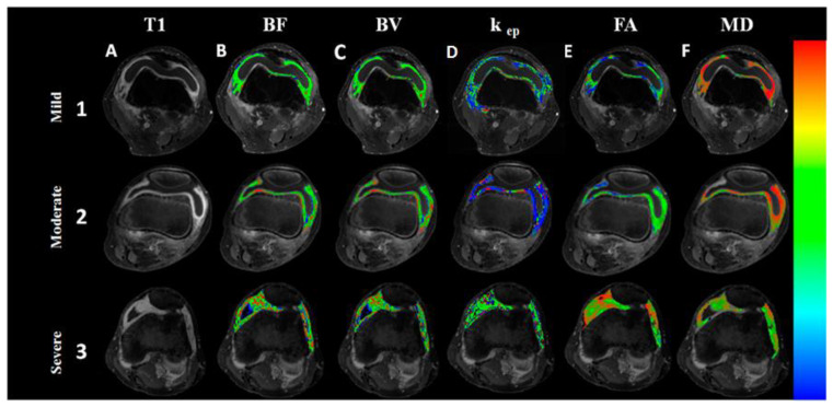
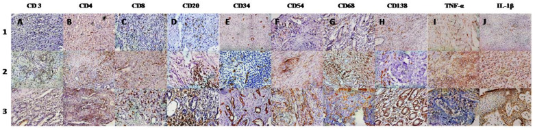
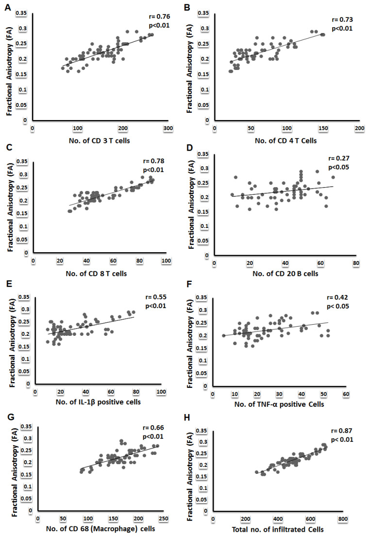
Objectives: It is difficult to capture the severity of synovial inflammation on imaging. Herein we hypothesize that diffusion tensor imaging (DTI) derived metrics may delineate the aggregation of the inflammatory cells and expression of inflammatory cytokines and dynamic contrast-enhanced (DCE) imaging may provide information regarding vascularity in the inflamed synovium. Patients and methods: Patients with knee arthritis (>3-months duration) underwent conventional (T2-weighted fast spin echo and spin echo T1-weighted images) as well as DTI and DCE MRI and thereafter arthroscopic guided synovial biopsy. DCE and DTI metrics were extracted from the masks of the segments of the inflamed synovium which enhanced on post-contrast T1-weighted MRI. These metrics were correlated with immunohistochemistry (IHC) parameters of inflammation on synovium. Statistical analysis: Pearson’s correlation was performed to study the relationship between DTI- and DCE-derived metrics, IHC parameters, and post-contrast signal intensity. Linear regression model was used to predict the values of IHC parameters using various DTI and DCE derived metrics as predictors. Results: There were 80 patients (52 male) with mean age 39.78 years and mean disease duration 19.82 months. Nineteen patients had tuberculosis and the rest had chronic undifferentiated monoarthritis (n = 31), undifferentiated spondyloarthropathy (n = 14), rheumatoid arthritis (n = 6), osteoarthritis (n = 4), reactive arthritis (n = 3), ankylosing spondylitis (n = 2), and juvenile idiopathic arthritis (n = 1). Fractional anisotropy (FA), a metric of DTI, had significant correlation with number of immune cells (r = 0.87, p < 0.01) infiltrating into the synovium and cytokines (IL-1β, r = 0.55, p < 0.01; TNF-α, r = 0.42, p < 0.01) in all patients and also in each group of patients and adhesion molecule expressed on these cells in all patients (CD54, r = 0.51, p < 0.01). DCE parameters significantly correlated with CD34 (blood flow, r = 0.78, p < 0.01; blood volume, r = 0.76, p < 0.01) in each group of patients, a marker of neo-angiogenesis. FA was the best predictor of infiltrating inflammatory cells, adhesion molecule and proinflammatory cytokines. Amongst the DCE parameters, blood volume, was best predictor of CD34. Conclusion: DTI and DCE metrics capture cellular and molecular markers of synovial inflammation in patients with chronic inflammatory arthritis.
Keywords: ankylosing spondylitis; markers of inflammation; osteoarthritis; rheumatoid arthritis; synovial histology; tuberculosis; undifferentiated arthritis.
Conflict of interest statement
The authors declare no conflict of interest.
Figures
Similar articles
-
Diffusion tensor anisotropy magnetic resonance imaging: a new tool to assess synovial inflammation.Rheumatology (Oxford). 2009 Apr;48(4):378-82. doi: 10.1093/rheumatology/ken499. Epub 2009 Jan 27. Rheumatology (Oxford). 2009. PMID: 19174567
-
Diffusion-weighted imaging for assessment of synovial inflammation in juvenile idiopathic arthritis: a promising imaging biomarker as an alternative to gadolinium-based contrast agents.Eur Radiol. 2017 Nov;27(11):4889-4899. doi: 10.1007/s00330-017-4876-y. Epub 2017 Jun 12. Eur Radiol. 2017. PMID: 28608162 Free PMC article.
-
Gadolinium-free assessment of synovitis using diffusion tensor imaging.NMR Biomed. 2022 Jan;35(1):e4614. doi: 10.1002/nbm.4614. Epub 2021 Sep 21. NMR Biomed. 2022. PMID: 34549476 Free PMC article.
-
Use of DCE-MRI in arthritis in the appendicular skeleton: a narrative review.Skeletal Radiol. 2025 Jun 10. doi: 10.1007/s00256-025-04966-7. Online ahead of print. Skeletal Radiol. 2025. PMID: 40495074 Review.
-
The role of diffusion tensor imaging and fractional anisotropy in the evaluation of patients with idiopathic normal pressure hydrocephalus: a literature review.Neurosurg Focus. 2016 Sep;41(3):E12. doi: 10.3171/2016.6.FOCUS16192. Neurosurg Focus. 2016. PMID: 27581308 Review.
Cited by
-
Magnetic Resonance Imaging (MRI)-Based Semi-Quantitative Methods for Rheumatoid Arthritis: From Scoring to Measurement.J Clin Med. 2024 Jul 16;13(14):4137. doi: 10.3390/jcm13144137. J Clin Med. 2024. PMID: 39064179 Free PMC article. Review.
-
Mobile health technologies in inflammatory bowel disease: a narrative review.BMC Gastroenterol. 2025 Aug 18;25(1):595. doi: 10.1186/s12876-025-04189-z. BMC Gastroenterol. 2025. PMID: 40826022 Free PMC article.
-
A Case Report of Intratesticular Hematoma in a Patient with Reiter's Syndrome.Diagnostics (Basel). 2023 Jun 7;13(12):1993. doi: 10.3390/diagnostics13121993. Diagnostics (Basel). 2023. PMID: 37370888 Free PMC article.
-
Evidence Suggesting Prolonged Neuroinflammation in a Subset of Children after Moderate/Severe TBI: A UCLA RAPBI Study.medRxiv [Preprint]. 2025 Jan 22:2025.01.20.25320782. doi: 10.1101/2025.01.20.25320782. medRxiv. 2025. PMID: 39974138 Free PMC article. Preprint.
-
Advanced Magnetic Resonance Imaging and Molecular Imaging of the Painful Knee.Semin Musculoskelet Radiol. 2023 Dec;27(6):618-631. doi: 10.1055/s-0043-1775741. Epub 2023 Nov 7. Semin Musculoskelet Radiol. 2023. PMID: 37935208 Free PMC article. Review.
References
-
- Melchiorri C., Meliconi R., Frizziero L., Silvestri T., Pulsatelli L., Mazzetti I., Borzì R.M., Uguccioni M., Facchini A. Enhanced and coordinated in vivo expression of inflammatory cytokines and nitric oxide synthase by chondrocytes from patients with osteoarthritis. Arthritis Rheum. 1998;41:2165–2174. doi: 10.1002/1529-0131(199812)41:12<2165::AID-ART11>3.0.CO;2-O. - DOI - PubMed
-
- Kirkham B.W., Lassere M.N., Edmonds J.P., Juhasz K.M., Bird P.A., Lee C.S., Shnier R., Portek I.J. Synovial membrane cytokine expression is predictive of joint damage progression in rheumatoid arthritis: A two-year prospective study (the DAMAGE study cohort) Arthritis Rheum. 2006;54:1122–1131. doi: 10.1002/art.21749. - DOI - PubMed
Grants and funding
LinkOut - more resources
Full Text Sources
Research Materials
