

Giant Bilateral Hydronephrosis in A Newborn-A Case Report
- PMID: 36553334
- PMCID: PMC9776467
- DOI: 10.3390/children9121890
Giant Bilateral Hydronephrosis in A Newborn-A Case Report
Abstract
Background: Prenatal hydronephrosis is common and may vary in size. Although mostly unproblematic, it may be a sign of urinary tract obstruction of differing severity.
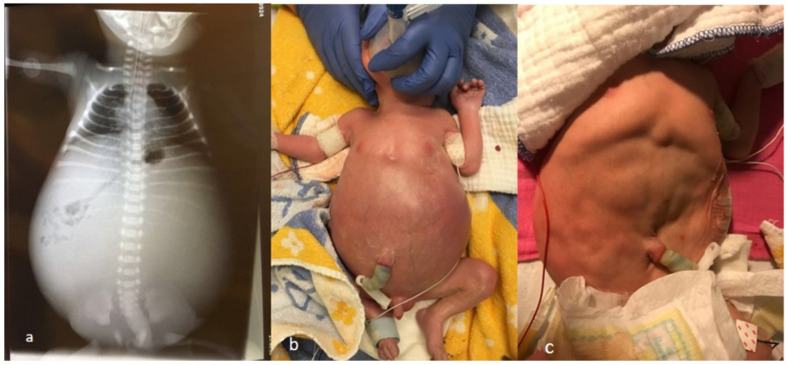
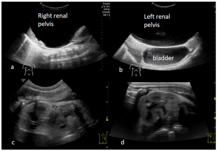
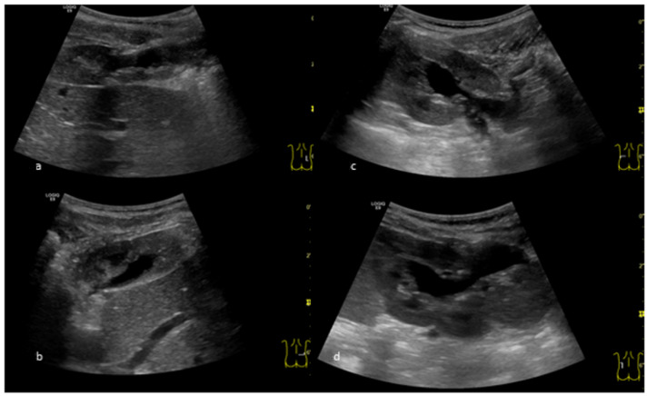
Case diagnosis/treatment: We present a boy with prenatally detected bilateral giant hydronephrosis. A prenatal ultrasound showed the whole abdominal cavity of the fetus filled with urine. Kidney parenchyma could not be seen. The boy was born at 34 + 1 weeks' gestation. After delivery, he showed a severely distended abdomen. Insertion of a nasogastric tube was not possible, and he had to be intubated due to respiratory distress. A bilateral percutaneous nephrostomy was performed immediately. After a few hours, he could be stabilized and extubated. An ultrasound on the following day showed two kidney units with normal kidney parenchyma of normal size. The initially slightly elevated serum creatinine level normalized within one week. An antegrade pyelography via the nephrostomy tubes showed bilateral ureteropelvic junction obstruction.
Conclusion: Severe bilateral hydronephrosis may be associated with good outcome and well-preserved kidney function. Prenatal counseling should be done carefully, with discussion of different treatment possibilities and without definitive prediction of outcome.
Keywords: CAKUT (congenital anomalies of the kidney and urinary tract); giant hydronephrosis; newborn; prenatal; ureteropelvic junction obstruction.
Conflict of interest statement
The authors declare no conflict of interest.
Figures
Similar articles
-
Long-term nephrostomy in an adult male spinal cord injury patient who had normal upper urinary tracts but developed bilateral hydronephrosis following penile sheath drainage: pyeloplasty and balloon dilatation of ureteropelvic junction proved futile: a case report.Cases J. 2009 Dec 16;2:9335. doi: 10.1186/1757-1626-2-9335. Cases J. 2009. PMID: 20062594 Free PMC article.
-
Outcome after prenatal diagnosis of congenital anomalies of the kidney and urinary tract.Eur J Pediatr. 2016 May;175(5):667-76. doi: 10.1007/s00431-015-2687-1. Epub 2016 Jan 25. Eur J Pediatr. 2016. PMID: 26805407
-
Improved split renal function after percutaneous nephrostomy in young adults with severe hydronephrosis due to ureteropelvic junction obstruction.J Urol. 2015 Jan;193(1):191-5. doi: 10.1016/j.juro.2014.07.005. Epub 2014 Jul 9. J Urol. 2015. PMID: 25014578
-
[Postnatal management of urinary tract anomalies after antenatal diagnosis].J Gynecol Obstet Biol Reprod (Paris). 2003 Jun;32(4):300-13. J Gynecol Obstet Biol Reprod (Paris). 2003. PMID: 12843878 Review. French.
-
Giant hydronephrosis due to ureteropelvic junction obstruction in a child: CT and MR appearances.Clin Imaging. 2002 Mar-Apr;26(2):125-8. doi: 10.1016/s0899-7071(01)00369-2. Clin Imaging. 2002. PMID: 11852221 Review.
Cited by
-
Missed diagnosis of giant hydronephrosis mimicking abdominal lymphatic malformation or omental cyst in a 16-year-old girl: A case report.Int J Surg Case Rep. 2025 Aug;133:111533. doi: 10.1016/j.ijscr.2025.111533. Epub 2025 Jun 19. Int J Surg Case Rep. 2025. PMID: 40570485 Free PMC article.
-
Pyeloureteral Junction Syndrome in a Neonate With a Solitary Kidney Treated by Anderson-Hynes Pyeloplasty: A Case Report.Cureus. 2024 Jul 28;16(7):e65589. doi: 10.7759/cureus.65589. eCollection 2024 Jul. Cureus. 2024. PMID: 39192924 Free PMC article.
References
Publication types
LinkOut - more resources
Full Text Sources
