

Early Regressive Development of the Subcommissural Organ of Two Human Fetuses with Non-Communicating Hydrocephalus
- PMID: 36553409
- PMCID: PMC9776597
- DOI: 10.3390/children9121966
Early Regressive Development of the Subcommissural Organ of Two Human Fetuses with Non-Communicating Hydrocephalus
Abstract
Hydrocephalus is a central nervous system condition characterized by CSF buildup and ventricular hypertrophy. It is divided into two types: communicative and non-communicating hydrocephalus. Congenital hydrocephalus has been linked to several changes in the subcommissural organ (SCO). However, it is unclear whether these changes occur before or as a result of the hydrocephalic illness. This report presents three cases of human fetuses with hydrocephalus: one non-communicating case, two communicating cases, and two controls. Hematoxylin-Eosin (H&E) or cresyl violet and immunohistochemistry with anti-transthyretin were used to analyze SCO morphological and secretory changes. We conclude that in the cases presented here, there could be an early regression in the SCO of the communicating cases that is not present in the non-communicating case.
Keywords: human congenital hydrocephalus; subcommissural organ; transthyretin.
Conflict of interest statement
The authors declare no conflict of interest.
Figures
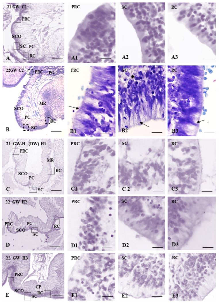
 ependymal cells,
  hypendymal cells.
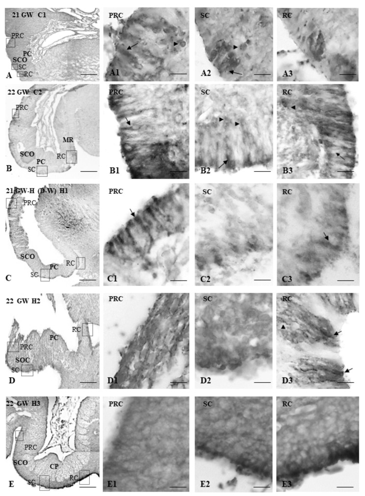
 ependymal cells, (▲) hypendymal cells. In the case of non-communicating hydrocephalus, TTR-ir can be observed in the rostral and caudal parts of the SCO. However, in the H2 case of communicating hydrocephalus, the TTR-ir can mainly be observed in the sub and retrocomisural parts of the SCO and located in the ependymal cells. In the H3 case of communicating the TTR-ir is in the ependymal cells of the retro-commissural parts, mainly in its apical pole.
Similar articles
-
The value of early and comprehensive diagnoses in a human fetus with hydrocephalus and progressive obliteration of the aqueduct of Sylvius: Case Report.BMC Neurol. 2016 Apr 11;16:45. doi: 10.1186/s12883-016-0566-7. BMC Neurol. 2016. PMID: 27067115 Free PMC article.
-
Reduced subcommissural organ glycoprotein immunoreactivity precedes aqueduct closure and ventricular dilatation in H-Tx rat hydrocephalus.Cell Tissue Res. 2004 Mar;315(3):361-73. doi: 10.1007/s00441-003-0843-9. Epub 2004 Jan 14. Cell Tissue Res. 2004. PMID: 14722750
-
Subcommissural organ, cerebrospinal fluid circulation, and hydrocephalus.Microsc Res Tech. 2001 Mar 1;52(5):591-607. doi: 10.1002/1097-0029(20010301)52:5<591::AID-JEMT1043>3.0.CO;2-7. Microsc Res Tech. 2001. PMID: 11241868 Review.
-
Reissner's fibre proteins and p73 variations in the cerebrospinal fluid and subcommissural organ of hydrocephalic rat.Anat Histol Embryol. 2009 Aug;38(4):282-5. doi: 10.1111/j.1439-0264.2009.00939.x. Epub 2009 Jun 10. Anat Histol Embryol. 2009. PMID: 19519738
-
Evidence of the subcommissural organ in humans and its association with hydrocephalus.Neurosurg Rev. 2002 Aug;25(4):205-15. doi: 10.1007/s10143-002-0208-y. Epub 2002 Apr 6. Neurosurg Rev. 2002. PMID: 12172724 Review.
References
-
- Höfer H. Zur Morphologie der circumventriculären Organe des Zwischenhirnes der Säugetiere. Zool Anz Suppl. 1959;22:202–251.
-
- Oksche A. Funktionelle histologische Untersuchungen über die Organe des Zwischenhirndaches der Chordaten. Anat. Anz. 1956;102:404–419. - PubMed
-
- Oksche A. Phylogenetic and conceptual aspects of the subcommissural organ. In: Oksche A., Rodríguez E.M., Fernández-Llébrez P., editors. The Subcommissural Organ, an Ependymal Brain Gland. Springer; Berlin/Heidelberg, Germany: 1993. pp. 23–32.
LinkOut - more resources
Full Text Sources
Research Materials
