Case Reports
. 2022 Nov 22;13(12):2184.
doi: 10.3390/genes13122184.

White Sponge Nevus Caused by Keratin 4 Gene Mutation: A Case Report

Affiliations
Case Reports

White Sponge Nevus Caused by Keratin 4 Gene Mutation: A Case Report

Yahui Qiao et al. Genes (Basel). .

Abstract

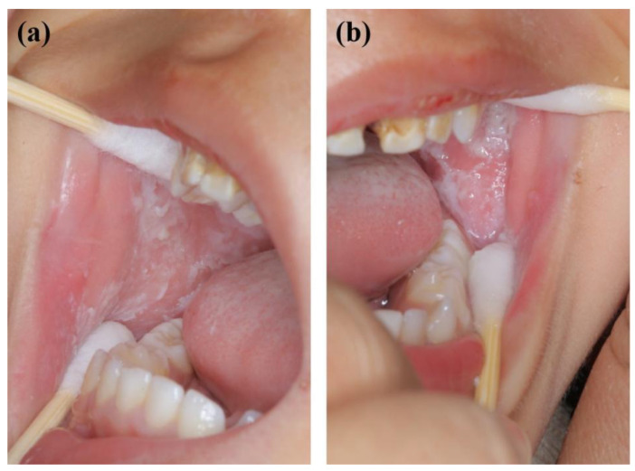
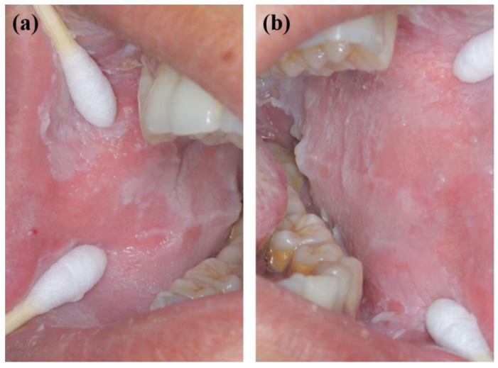
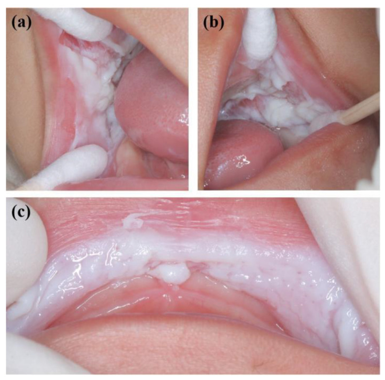
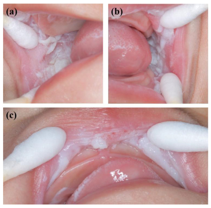
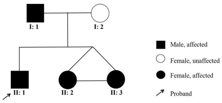
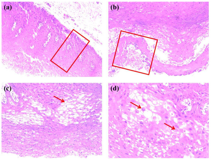
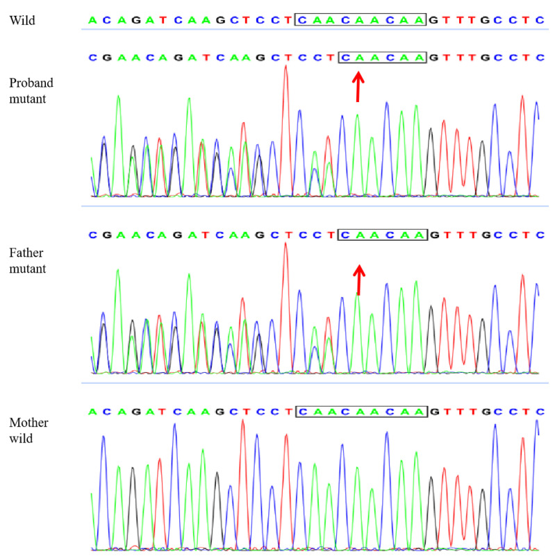
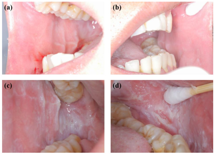
White sponge nevus (WSN) is a rare autosomal dominant disease with a family history, often caused by mutations of the keratin 4 (K4) and keratin 13 (K13) genes in patients. It is characterized by frequently occurred white corrugated folds in the bilateral buccal mucosa with soft texture. On histopathological examination, hyperkeratosis of epithelial cells, edema, and vacuolar changes in the spinous cells are observed in the lesions, despite a normal layer of basal cells. WSN should be differentiated from other oral white spot diseases, mainly oral lichen planus, oral candidiasis, oral white edema, and Heck's disease, to reduce misdiagnosis and unnecessary treatment. At present, there is no specific treatment method. The purpose of this study was to report the clinical data of four WSN patients of the same family with the K4 gene mutation. The occurrence of WSN in a pair of monozygotic twins with very similar clinical presentations was identified for the first time. The gene sequencing results showed that there was a heterozygous deletion (C. 438_440delCAA) in exon 1 of the K4 gene, resulting in an aspartic acid loss in both the proband and his father. Finally, the etiology, pathogenesis, pathological manifestations, clinical manifestations, diagnosis, differential diagnosis, and related treatment methods are discussed to provide a reference for clinical treatment of the disease.

Keywords: K4; WSN; gene mutation; genetic disorder; recurrence.


Conflict of interest statement

The authors declare no conflict of interest.

Figures

Figure 1
Figure 1
Oral manifestations of the proband (the 2-year-old boy) showing large gray and white folds and plaques in the (a) right and (b) left buccal mucosa.
Figure 2
Figure 2
Oral manifestations of the father showing large gray and white folds and plaques in the right (a) and left (b) buccal mucosa.
Figure 3
Figure 3
Oral manifestations of sister 1 showing large white corrugated folds in the (a) right and (b) left buccal mucosa and (c) inner mucosa of the upper lip, resembling a sponge.
Figure 4
Figure 4
Oral manifestations of sister 2 showing large white corrugated folds in the (a) right and (b) left buccal mucosa and (c) inner mucosa of the upper lip, resembling a sponge.
Figure 5
Figure 5
Pedigree of the proband’s family.
Figure 6
Figure 6
Hematoxylin-eosin staining of the right buccal mucosa of the proband’s father with (a) the red frame indicating enlarged spinous cells and well differentiated basal cells with increased number of layers (40×), (b) the red frame indicating incomplete keratinization of surface cells and uneven epithelial tissue with no inflammatory infiltration (100×), (c) the red arrow indicating vacuolar change of spinous cells (200×), and (d) the top and bottom arrows indicating spinous cells with nuclear pyknosis and cell edema, respectively (400×).
Figure 7
Figure 7
Gene sequencing results showing the heterozygous deletion (C. 438_440delCAA) in exon 1 of the K4 gene in the proband and his father, namely the aspartic acid loss, and no abnormality in the mother, with the arrows indicating the missing bases.
Figure 8
Figure 8
Oral mucosal examination for the proband’s father at follow-up showing significant reduction of lesions in the (a) right and (b) left buccal mucosa and a nearly complete disappearance of white corrugated plaques after one week and recurrence in the (c) right and (d) left buccal mucosa with greyish white corrugated folds and plaques after three months.

Similar articles

Cited by

References

    1. Sanjeeta N., Nandini D.B., Premlata T., Banerjee S. White sponge nevus: Report of three cases in a single family. J. Oral Maxillofac. Pathol. 2016;20:300–303. doi: 10.4103/0973-029X.185915. - DOI - PMC - PubMed
    1. Akimori T., Ueda D., Takada M., Kabata K., Fukui S., Miyawaki A. A case of non-hereditary white sponge nevus in the oral cavity successfully treated with tetracycline ointment and oral multivitamins. J. UOEH. 2021;43:421–426. doi: 10.7888/juoeh.43.421. - DOI - PubMed
    1. Chen Q.M. Oral Mucosal Pathology. 5th ed. People’s Medical Publishing House; Beijing, China: 2020. pp. 107–108.
    1. Moll R., Divo M., Langbein L. The human keratins: Biology and pathology. Histochem. Cell Biol. 2008;129:705–733. doi: 10.1007/s00418-008-0435-6. - DOI - PMC - PubMed
    1. Cai W., Jiang B., Yu F., Yang J., Chen Z., Liu J., Wei R., Zhao S., Wang X., Liu S. Current approaches to the diagnosis and treatment of white sponge nevus. Expert Rev. Mol. Med. 2015;17:e9. doi: 10.1017/erm.2015.7. - DOI - PubMed

Publication types