

SLC26A4 Phenotypic Variability Influences Intra- and Inter-Familial Diagnosis and Management
- PMID: 36553459
- PMCID: PMC9778369
- DOI: 10.3390/genes13122192
SLC26A4 Phenotypic Variability Influences Intra- and Inter-Familial Diagnosis and Management
Abstract
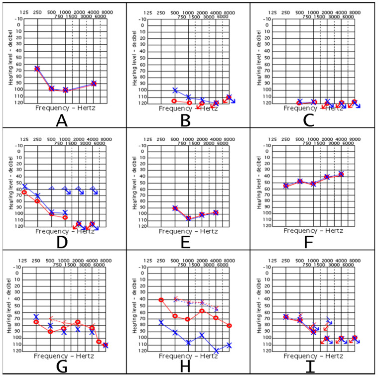
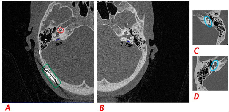
SLC26A4 is one of the most common genes causing autosomal recessive non-syndromic sensorineural hearing loss (SNHL). It has been reported to cause Pendred Syndrome (PDS) and DFNB4 which is deafness with enlarged vestibular aqueduct (EVA). However, mutated SLC26A4 is not conclusive for having either DFNB4 or PDS. Three unrelated Jordanian families consisting of eight affected individuals with congenital bilateral hearing loss (HL) participated in this study. Whole-exome and Sanger sequencing were performed to investigate the underlying molecular etiology of HL. Further clinical investigations, including laboratory blood workup for the thyroid gland, CT scan for the temporal bone, and thyroid ultrasound were performed. Three disease-causing variants were identified in SLC26A4 in the three families, two of which were novel. Two families had a novel pathogenic homozygous splice-site accepter variant (c.165-1G>C), while the third family had compound heterozygous pathogenic variants (c.1446G>A; p.Trp482* and c.304G>A; p.Gly102Arg). Our approach helped in redirecting the diagnosis of several affected members of three different families from non-syndromic HL to syndromic HL. Two of the affected individuals had typical PDS, one had DFNB4, while the rest had atypical PDS. Our work emphasized the intra- and inter-familial variability of SLC26A4-related phenotypes. In addition, we highlighted the variable phenotypic impact of SLC26A4 on tailoring a personalized healthcare management.
Keywords: DFNB4; Pendred syndrome; SLC26A4; enlarged vestibular aqueduct; hearing loss; heterogeneity.
Conflict of interest statement
The authors declare no conflict of interest.
Figures


Similar articles
-
Analysis of SLC26A4, FOXI1, and KCNJ10 Gene Variants in Patients with Incomplete Partition of the Cochlea and Enlarged Vestibular Aqueduct (EVA) Anomalies.Int J Mol Sci. 2022 Dec 6;23(23):15372. doi: 10.3390/ijms232315372. Int J Mol Sci. 2022. PMID: 36499699 Free PMC article.
-
Genetic heterogeneity in patients with enlarged vestibular aqueduct and Pendred syndrome.Mol Med. 2025 May 27;31(1):208. doi: 10.1186/s10020-025-01262-x. Mol Med. 2025. PMID: 40426046 Free PMC article.
-
Novel genetic determinants contribute to hearing loss in a central European cohort with enlarged vestibular aqueduct.Mol Med. 2025 Mar 22;31(1):111. doi: 10.1186/s10020-025-01159-9. Mol Med. 2025. PMID: 40121402 Free PMC article.
-
Genetic architecture and phenotypic landscape of SLC26A4-related hearing loss.Hum Genet. 2022 Apr;141(3-4):455-464. doi: 10.1007/s00439-021-02311-1. Epub 2021 Aug 3. Hum Genet. 2022. PMID: 34345941 Review.
-
Variability in Inner Ear Morphology Among a Family With Pendred Syndrome Due to a SLC26A4 Gene Variant.Ann Otol Rhinol Laryngol. 2024 Sep;133(9):828-833. doi: 10.1177/00034894241261491. Epub 2024 Jun 14. Ann Otol Rhinol Laryngol. 2024. PMID: 38877731 Review.
Cited by
-
Unraveling the genetic tapestry of pediatric sarcomeric cardiomyopathies and masquerading phenocopies in Jordan.Sci Rep. 2024 Jul 2;14(1):15141. doi: 10.1038/s41598-024-64921-9. Sci Rep. 2024. PMID: 38956129 Free PMC article.
References
-
- Royaux I.E., Suzuki K., Mori A., Katoh R., Everett L.A., Kohn L.D., Green E.D. Pendrin, the Protein Encoded by the Pendred Syndrome Gene (PDS), Is an Apical Porter of Iodide in the Thyroid and Is Regulated by Thyroglobulin in FRTL-5 Cells. Endocrinology. 2000;141:839–845. doi: 10.1210/endo.141.2.7303. - DOI - PubMed
-
- Royaux I.E., Wall S.M., Karniski L.P., Everett L.A., Suzuki K., Knepper M.A., Green E.D. Pendrin, Encoded by the Pendred Syndrome Gene, Resides in the Apical Region of Renal Intercalated Cells and Mediates Bicarbonate Secretion. Proc. Natl. Acad. Sci. USA. 2001;98:4221–4226. doi: 10.1073/pnas.071516798. - DOI - PMC - PubMed
-
- Park H., Shaukat S., Liu X., Hahn S.H., Naz S., Ghosh M., Kim H., Moon S., Abe S., Tukamoto K., et al. Origins and Frequencies of SLC26A4 (PDS) Mutations in East and South Asians: Global Implications for the Epidemiology of Deafness. J. Med. Genet. 2003;4:242–249. doi: 10.1136/jmg.40.4.242. - DOI - PMC - PubMed
Publication types
MeSH terms
Substances
Supplementary concepts
LinkOut - more resources
Full Text Sources
Miscellaneous
