. 2022 Dec 13;13(12):2348.
doi: 10.3390/genes13122348.

Challenges in Gene Therapy for Somatic Reverted Mosaicism in X-Linked Combined Immunodeficiency by CRISPR/Cas9 and Prime Editing

Affiliations

Challenges in Gene Therapy for Somatic Reverted Mosaicism in X-Linked Combined Immunodeficiency by CRISPR/Cas9 and Prime Editing

Yujuan Hou et al. Genes (Basel). .

Abstract

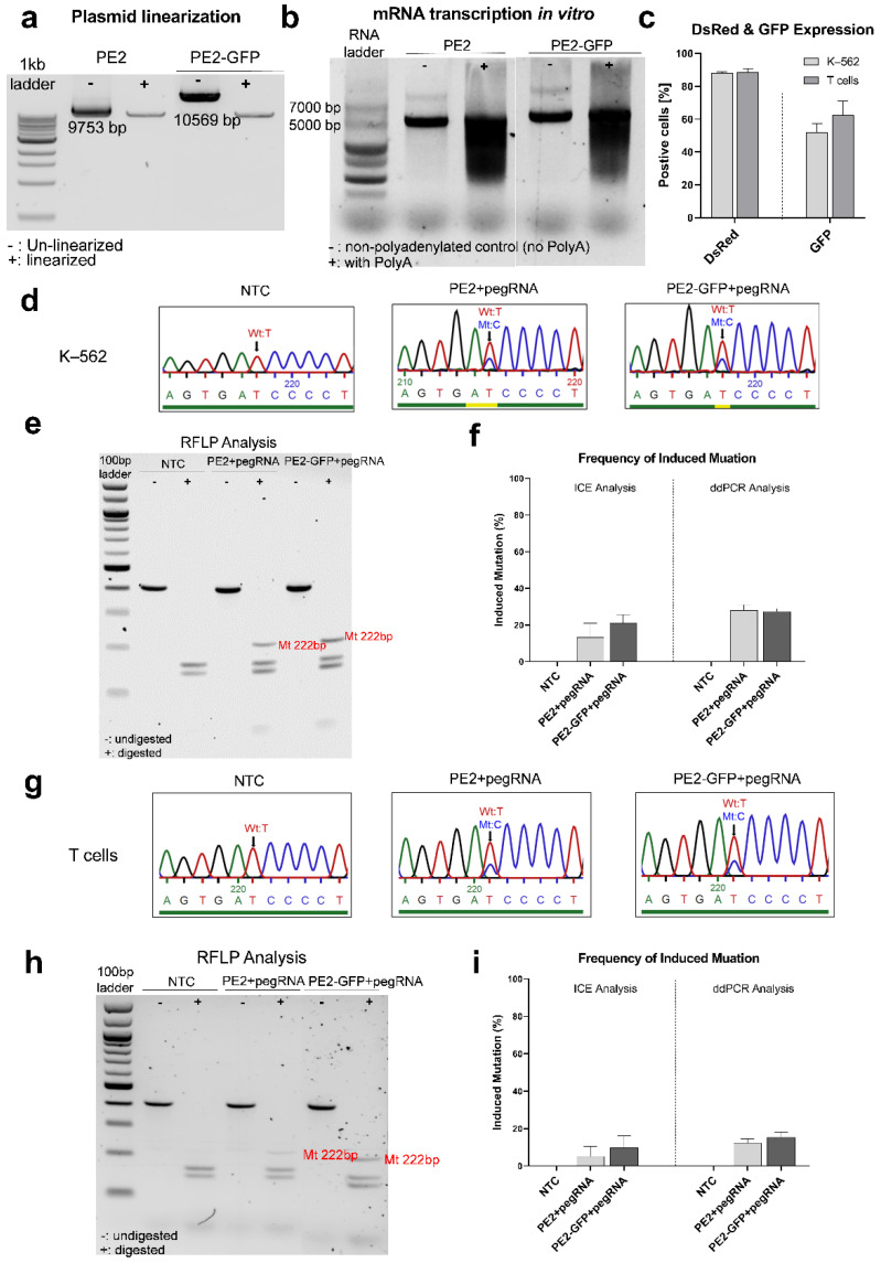
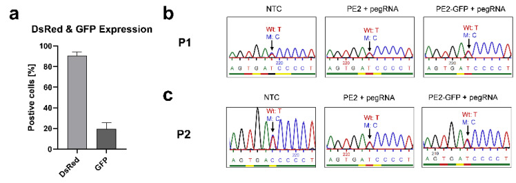
X-linked severe combined immunodeficiency (X-SCID) is a primary immunodeficiency that is caused by mutations in the interleukin-2 receptor gamma (IL2RG) gene. Some patients present atypical X-SCID with mild clinical symptoms due to somatic revertant mosaicism. CRISPR/Cas9 and prime editing are two advanced genome editing tools that paved the way for treating immune deficiency diseases. Prime editing overcomes the limitations of the CRISPR/Cas9 system, as it does not need to induce double-strand breaks (DSBs) or exogenous donor DNA templates to modify the genome. Here, we applied CRISPR/Cas9 with single-stranded oligodeoxynucleotides (ssODNs) and prime editing methods to generate an in vitro model of the disease in K-562 cells and healthy donors' T cells for the c. 458T>C point mutation in the IL2RG gene, which also resulted in a useful way to optimize the gene correction approach for subsequent experiments in patients' cells. Both methods proved to be successful and were able to induce the mutation of up to 31% of treated K-562 cells and 26% of treated T cells. We also applied similar strategies to correct the IL2RG c. 458T>C mutation in patient T cells that carry the mutation with revertant somatic mosaicism. However, both methods failed to increase the frequency of the wild-type sequence in the mosaic T cells of patients due to limited in vitro proliferation of mutant cells and the presence of somatic reversion. To the best of our knowledge, this is the first attempt to treat mosaic cells from atypical X-SCID patients employing CRISPR/Cas9 and prime editing. We showed that prime editing can be applied to the formation of specific-point IL2RG mutations without inducing nonspecific on-target modifications. We hypothesize that the feasibility of the nucleotide substitution of the IL2RG gene using gene therapy, especially prime editing, could provide an alternative strategy to treat X-SCID patients without revertant mutations, and further technological improvements need to be developed to correct somatic mosaicism mutations.

Keywords: CRISPR/Cas9; IL2RG; SCID; prime editing; somatic mosaicism; ssODN.


Conflict of interest statement

Alicia Roig-Merino is employed at MaxCyte Inc. The authors declare no conflict of interest.

Figures

Figure 1
Figure 1
Frequency of the IL2RG c.458T>C mutation in mosaic T cells. Expanded CD3+ T cells of P1 were harvested to purify gDNA on days 1 and 5 post-isolation, separately. Sanger sequencing showed the frequency of the mutant (M) base, C, was 64% on day 1 and 47% on day 5 in the mosaic T cells (the blue, red, green, and black peaks stand in place of bases C, T, A, and G respectively).
Figure 2
Figure 2
Design and screening of sgRNAs. (a) Schematic representations of the IL2RG gene and designed sgRNAs were created in the Exon-Intron Graphic Maker (http://wormweb.org/exonintron) and modified in PowerPoint. the IL2RG gene’s eight exons are depicted: the c.458T>C mutation is presented on Exon4. The cutting sites of all designed sgRNAs are highlighted. sgRNA1 and 5 target the wild-type sequence; sgRNA3 and 4 target the mutant sequence; and sgRNA2 and 6 target both sequences. (b) In vitro CRISPR/Cas9 cutting assay. The visualization of the agarose gel showed that the PCR products (409 bp) were cut into fragments of different sizes after RNP incubation for 2 h (bands with 246 bp and 163 bp for sgRNA1; 251 bp and 158 bp for sgRNA2; 250 bp and 159 bp for sgRNA3; 243 bp and 166 bp for sgRNA4; 243 bp and 166 bp for sgRNA5; 364 bp and 45 bp for sgRNA6). sgRNA1, 2, 4, 5, and 6 similarly cut the target DNA amplicon of both the healthy donor (HD) and P1, while sgRNA3 only worked for the DNA of P1. (c) sgRNA screening of K–562 cells (n = 2). All sgRNAs led to a more than 70% indel frequency, except sgRNA3 (4%). (d) sgRNA screening of T cells in healthy donors (n = 3). sgRNA3 generated a 21% indel rate, whereas the rest of the sgRNAs generated a more than 60% indel rate. (e) sgRNA screening of T cells in patients (n = 2, P1 and P2). sgRNAs induced a 32–64% indel formation. (f) DsRed protein expression was detected 24 h post-transfection with flow cytometry as 90% in K–562 and T cells. (g) The proliferation rate of transfected T cells (healthy donors, n = 3) is calculated according to the fold-change (ratio) of the cell number between day 5 and day 1 post-electroporation. Compared with non-transfected cells, the proliferation rate was reduced significantly in cells transfected with IL2RG sgRNAs and Cas9 (****, p < 0.0001; ordinary one-way ANOVA test) but not in cells transfected with TRAC sgRNA and Cas9 (ns, p > 0.05; ordinary one-way ANOVA test). Mean ± SEM of biologically independent experiments is shown.
Figure 3
Figure 3
CRISPR/Cas9-ssODN transfection to induce mutation in K–562 cells and T cells from healthy donors. The transfection of RNP (IL2RG sgRNA2 and Cas9)-ssODN2 to induce IL2RG mutation in the K–562 cells (n = 2) and T cells of healthy donors (n = 3) was performed with MaxCyte®. (a) DsRed was expressed in more than 96% of K–562 cell and T cell control samples. (b) The indel frequencies were 96–97% (RNP transfection) and 78–84% (RNP-ssODN transfection) in K–562 cells and 66–80% (RNP transfection) and 35–54% (RNP-ssODN transfection) in T cells. Sanger sequencing results of (c) K–562 cells and (d) T cells transfected with RNP-ssODN showed induced mutant nucleotides of c.458C (targeting inducing mutant base) and c.459A (silent blocking mutation) (the blue, red, green, and black peaks stand in place of bases C, T, A, and G respectively). (e) HDR efficiency quantified by ICE analysis based on Sanger sequencing showed 3.5 ± 1.5% HDR in K–562 cells and 15.0 ± 5.7% HDR in T cells. Mean ± SEM of biologically independent experiments is shown.
Figure 4
Figure 4
CRISPR/Cas9-ssODN transfection in mosaic T cells to correct the IL2RG c.458T>C mutation. T cells of patients (n = 2, P1 and P2) were transfected with RNP (IL2RG sgRNA3/4 and Cas9)-ssODN3/4 to correct the c.458T>C IL2RG mutation with MaxCyte®. (a) DsRed mRNA was expressed in up to 90% of cells. (b) The indel frequency of edited samples with different RNPs and ssODNs was 26–72%. Mean ± SEM of biologically independent experiments is shown. The Sanger sequencing of non-transfected and transfected T cells with the Cas9, sgRNA4, and ssODN3/4 T cells of (c) P1 and (d) P2. (The blue, red, green, and black peaks stand in place of bases C, T, A, and G respectively). Compared with the unedited cells, the indel generation was visible, but no higher frequency of the wild-type nucleotide was observed in the edited samples.
Figure 5
Figure 5
Prime editing in K-562 cells and healthy donors’ T cells to induce the IL2RG c.458T>C mutation. Gel visualization of (a) linearized plasmids and (b) in vitro synthesized mRNAs. Inducing the IL2RG c.458T>C mutation in K–562 cells (n = 2) and healthy donors’ T cells (n = 3) was carried out using prime editing with PE2 and PE2-GFP mRNA and pegRNA1. The DsRed and GFP expressions, as transfection controls, were evaluated 24 h after electroporation. (c) Up to 88% of the K–562 cells and 91% of the T cells expressed DsRed; 41–62% of the K–562 cells and 47–76% of the T cells expressed GFP. The gDNA was acquired for a genomic analysis of editing efficiency two days post-electroporation. In K–562 cells, (d) PE2 mRNA-pegRNA and PE2-GFP mRNA-pegRNA-transfected cells showed the induced mutant base c.458C (the blue, red, green, and black peaks stand in place of bases C, T, A, and G respectively); (e) an RFLP analysis showed that a band with the expected size (222 bp) after the digestion of a PCR product (409 bp) by the DpnII enzyme for the mutant sequence in the PE-pegRNA-transfected samples. (f) The frequency of induced mutation was evaluated using ICE and ddPCR analysis: 26.5 ± 2.5% (PE2 mRNA-pegRNA) and 29.0 ± 1.0% (PE2-GFP mRNA-pegRNA) via ICE analysis; 28.0 ± 3.0% (PE2 mRNA-pegRNA) and 27.5 ± 1.5% (PE2-GFP mRNA-pegRNA) via ddPCR analysis. In the T cells of healthy donors, (g) PE2 mRNA-pegRNA and PE2-GFP mRNA-pegRNA transfection samples showed the induced mutant base c.458C; (h) an RFLP analysis revealed mutant generation in the edited samples with PE-pegRNA. (i) The frequency of induced mutation was 16.7 ± 8.4% (PE2 mRNA-pegRNA) and 21.0 ± 4.0% (PE2-GFP mRNA-pegRNA) via ICE analysis and 13.1 ± 3.0% (PE2 mRNA-pegRNA) and 18.0 ± 5.3% (PE2-GFP mRNA-pegRNA) via ddPCR analysis. Mean ± SEM of biologically independent experiments are provided.
Figure 6
Figure 6
Prime editing in mosaic T cells to correct IL2RG c.458T>C mutation. PE (PE2/PE2-GFP mRNA)–pegRNA2 (carrying wild-type base c.458T of IL2RG) was transfected to edit the IL2RG c.458T>C mutation in the mosaic T cells of the patients (n = 2, P1 and P2). (a) Up to 92% and 19% of cells expressed DsRed and GFP, respectively. Sanger sequencing did not exhibit significate differences between the NTC (non-transfected control) and edited samples (transfected with PE2/PE2-GFP mRNA-pegRNA) in either (b) P1 and (c) P2 (the blue, red, green, and black peaks stand in place of bases C, T, A, and G respectively). Mean ± SEM of biologically independent experiments is shown.

Similar articles

Cited by

References

    1. Puck J.M., Deschênes S.M., Porter J.C., Dutra A.S., Brown C.J., Willard H.F., Henthorn P.S. The interleukin-2 receptor γ chain maps to Xq13.1 and is mutated in X-linked severe combined immunodeficiency, SCIDX1. Hum. Mol. Genet. 1993;2:1099–1104. doi: 10.1093/hmg/2.8.1099. - DOI - PubMed
    1. Gaspar H.B., Qasim W., Davies E.G., Rao K., Amrolia P.J., Veys P. How I treat severe combined immunodeficiency. Blood. 2013;122:3749–3758. doi: 10.1182/blood-2013-02-380105. - DOI - PubMed
    1. Kumrah R., Vignesh P., Patra P., Singh A., Anjani G., Saini P., Sharma M., Kaur A., Rawat A. Genetics of severe combined immunodeficiency. Genes Dis. 2020;7:52–61. doi: 10.1016/j.gendis.2019.07.004. - DOI - PMC - PubMed
    1. Lim C.K., Abolhassani H., Appelberg S.K., Sundin M., Hammarström L. hypomorphic mutation: Identification of a novel pathogenic mutation in exon 8 and a review of the literature. Allergy Asthma Clin. Immunol. Off. J. Can. Soc. Allergy Clin. Immunol. 2019;15:2. doi: 10.1186/s13223-018-0317-y. - DOI - PMC - PubMed
    1. Stephan V., Wahn V., Le Deist F., Dirksen U., Broker B., Müller-Fleckenstein I., Horneff G., Schroten H., Fischer A., de Saint Basile G. Atypical X-linked severe combined immunodeficiency due to possible spontaneous reversion of the genetic defect in T cells. N. Engl. J. Med. 1996;335:1563–1567. doi: 10.1056/NEJM199611213352104. - DOI - PubMed

Publication types

MeSH terms

Supplementary concepts

LinkOut - more resources