

Clinical Decision Support Framework for Segmentation and Classification of Brain Tumor MRIs Using a U-Net and DCNN Cascaded Learning Algorithm
- PMID: 36553864
- PMCID: PMC9777942
- DOI: 10.3390/healthcare10122340
Clinical Decision Support Framework for Segmentation and Classification of Brain Tumor MRIs Using a U-Net and DCNN Cascaded Learning Algorithm
Abstract
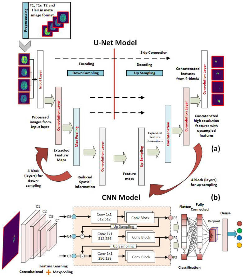
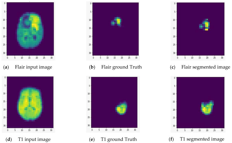
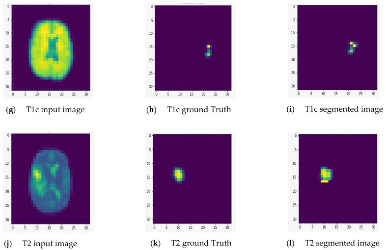
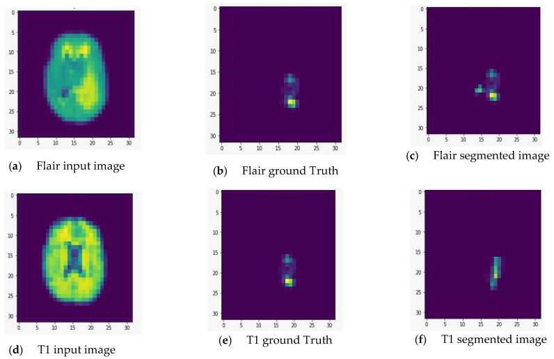
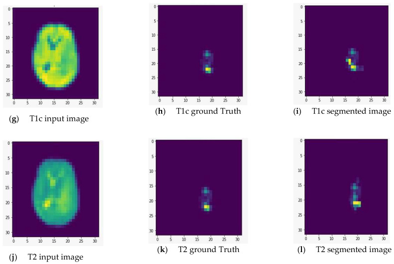
Brain tumors (BTs) are an uncommon but fatal kind of cancer. Therefore, the development of computer-aided diagnosis (CAD) systems for classifying brain tumors in magnetic resonance imaging (MRI) has been the subject of many research papers so far. However, research in this sector is still in its early stage. The ultimate goal of this research is to develop a lightweight effective implementation of the U-Net deep network for use in performing exact real-time segmentation. Moreover, a simplified deep convolutional neural network (DCNN) architecture for the BT classification is presented for automatic feature extraction and classification of the segmented regions of interest (ROIs). Five convolutional layers, rectified linear unit, normalization, and max-pooling layers make up the DCNN's proposed simplified architecture. The introduced method was verified on multimodal brain tumor segmentation (BRATS 2015) datasets. Our experimental results on BRATS 2015 acquired Dice similarity coefficient (DSC) scores, sensitivity, and classification accuracy of 88.8%, 89.4%, and 88.6% for high-grade gliomas. When it comes to segmenting BRATS 2015 BT images, the performance of our proposed CAD framework is on par with existing state-of-the-art methods. However, the accuracy achieved in this study for the classification of BT images has improved upon the accuracy reported in prior studies. Image classification accuracy for BRATS 2015 BT has been improved from 88% to 88.6%.
Keywords: CAD system; CNN; U-Net; brain tumor; classification; clinical decision; segmentation.
Conflict of interest statement
The authors declare no conflict of interest.
Figures
Similar articles
-
A Novel Approach for Fully Automatic Intra-Tumor Segmentation With 3D U-Net Architecture for Gliomas.Front Comput Neurosci. 2020 Feb 18;14:10. doi: 10.3389/fncom.2020.00010. eCollection 2020. Front Comput Neurosci. 2020. PMID: 32132913 Free PMC article.
-
Automatic Semantic Segmentation of Brain Gliomas from MRI Images Using a Deep Cascaded Neural Network.J Healthc Eng. 2018 Mar 19;2018:4940593. doi: 10.1155/2018/4940593. eCollection 2018. J Healthc Eng. 2018. PMID: 29755716 Free PMC article.
-
Brain tumor segmentation in MRI images using nonparametric localization and enhancement methods with U-net.Int J Comput Assist Radiol Surg. 2022 Mar;17(3):589-600. doi: 10.1007/s11548-022-02566-7. Epub 2022 Jan 29. Int J Comput Assist Radiol Surg. 2022. PMID: 35092598
-
The Application of Deep Convolutional Neural Networks to Brain Cancer Images: A Survey.J Pers Med. 2020 Nov 12;10(4):224. doi: 10.3390/jpm10040224. J Pers Med. 2020. PMID: 33198332 Free PMC article. Review.
-
Deep Learning in Selected Cancers' Image Analysis-A Survey.J Imaging. 2020 Nov 10;6(11):121. doi: 10.3390/jimaging6110121. J Imaging. 2020. PMID: 34460565 Free PMC article. Review.
Cited by
-
A New Breakpoint to Classify 3D Voxels in MRI: A Space Transform Strategy with 3t2FTS-v2 and Its Application for ResNet50-Based Categorization of Brain Tumors.Bioengineering (Basel). 2023 May 23;10(6):629. doi: 10.3390/bioengineering10060629. Bioengineering (Basel). 2023. PMID: 37370560 Free PMC article.
-
Application of Deep Learning for Prediction of Alzheimer's Disease in PET/MR Imaging.Bioengineering (Basel). 2023 Sep 24;10(10):1120. doi: 10.3390/bioengineering10101120. Bioengineering (Basel). 2023. PMID: 37892850 Free PMC article. Review.
-
Leveraging transfer learning-driven convolutional neural network-based semantic segmentation model for medical image analysis using MRI images.Sci Rep. 2024 Dec 18;14(1):30549. doi: 10.1038/s41598-024-81966-y. Sci Rep. 2024. PMID: 39695183 Free PMC article.
-
Advances in the Use of Deep Learning for the Analysis of Magnetic Resonance Image in Neuro-Oncology.Cancers (Basel). 2024 Jan 10;16(2):300. doi: 10.3390/cancers16020300. Cancers (Basel). 2024. PMID: 38254790 Free PMC article. Review.
-
Bridging the Gap: Exploring Opportunities, Challenges, and Problems in Integrating Assistive Technologies, Robotics, and Automated Machines into the Health Domain.Healthcare (Basel). 2023 Sep 4;11(17):2462. doi: 10.3390/healthcare11172462. Healthcare (Basel). 2023. PMID: 37685498 Free PMC article.
References
-
- Tian S., Yang W., Le Grange J.M., Wang P., Huang W., Ye Z. Smart Healthcare: Making Medical Care More Intelligent. Glob. Health J. 2019;3:62–65. doi: 10.1016/j.glohj.2019.07.001. - DOI
-
- Gong F.F., Sun X.Z., Lin J., Gu X.D. Primary Exploration in Establishment of China’s Intelligent Medical Treatment. Mod. Hosp. Manag. 2013;11:28–29.
-
- Farahani B., Firouzi F., Chang V., Badaroglu M., Constant N., Mankodiya K. Towards Fog-Driven IoT EHealth: Promises and Challenges of IoT in Medicine and Healthcare. Future Gener. Comput. Syst. 2018;78:659–676. doi: 10.1016/j.future.2017.04.036. - DOI
Grants and funding
LinkOut - more resources
Full Text Sources
Miscellaneous
