

Glaucoma Detection and Classification Using Improved U-Net Deep Learning Model
- PMID: 36554021
- PMCID: PMC9778546
- DOI: 10.3390/healthcare10122497
Glaucoma Detection and Classification Using Improved U-Net Deep Learning Model
Abstract
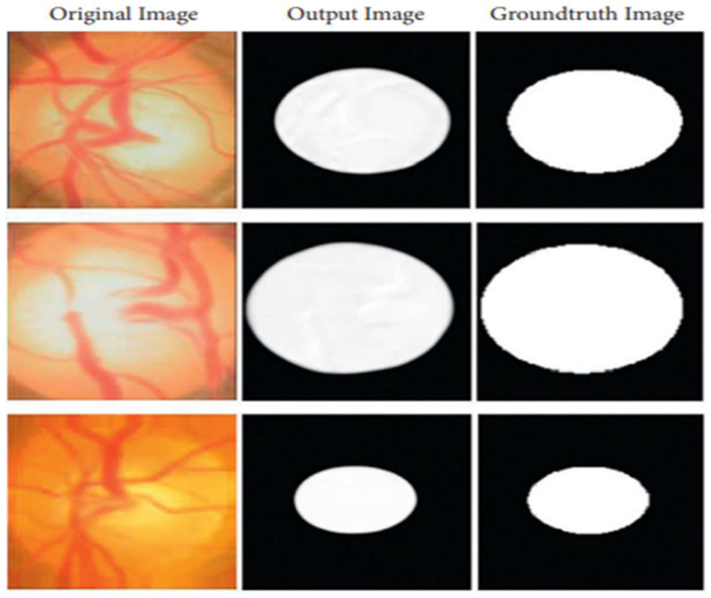
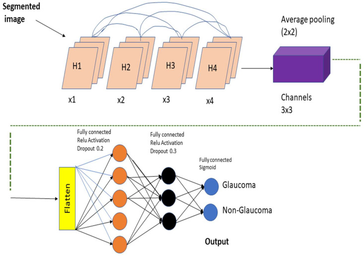
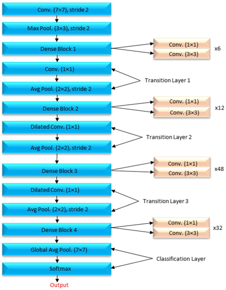
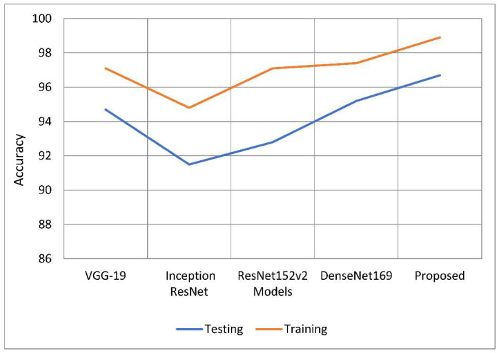
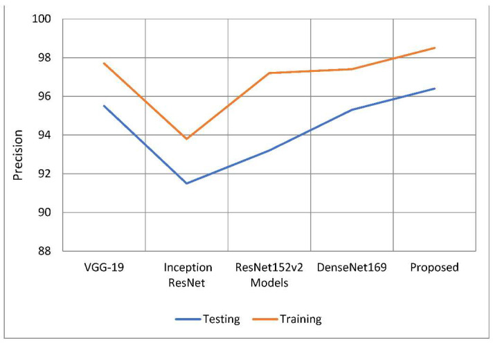
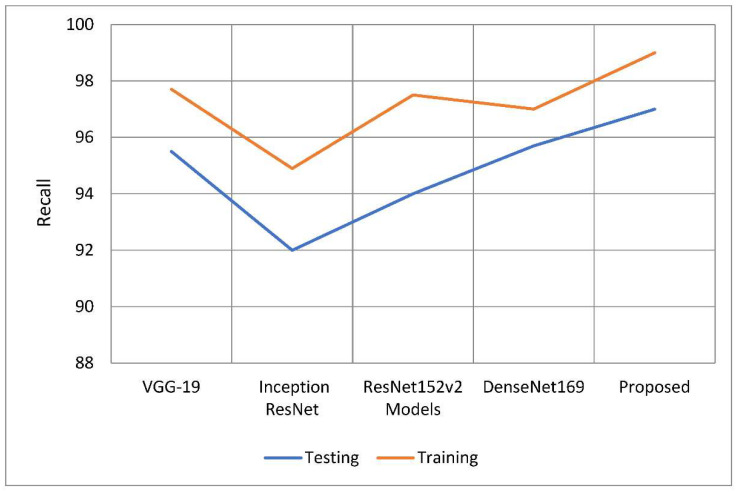
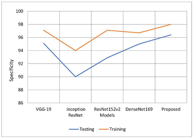
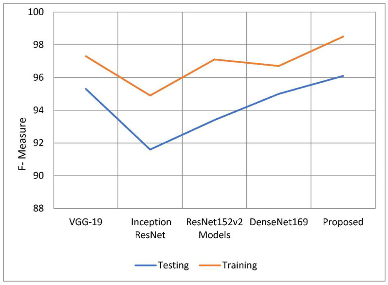
Glaucoma is prominent in a variety of nations, with the United States and Europe being two of the most famous. Glaucoma now affects around 78 million people throughout the world (2020). By the year 2040, it is expected that there will be 111.8 million cases of glaucoma worldwide. In countries that are still building enough healthcare infrastructure to cope with glaucoma, the ailment is misdiagnosed nine times out of ten. To aid in the early diagnosis of glaucoma, the creation of a detection system is necessary. In this work, the researchers propose using a technology known as deep learning to identify and predict glaucoma before symptoms appear. The glaucoma dataset is used in this deep learning algorithm that has been proposed for analyzing glaucoma images. To get the required results when using deep learning principles for the job of segmenting the optic cup, pretrained transfer learning models are integrated with the U-Net architecture. For feature extraction, the DenseNet-201 deep convolution neural network (DCNN) is used. The DCNN approach is used to determine whether a person has glaucoma. The fundamental goal of this line of research is to recognize glaucoma in retinal fundus images, which will aid in assessing whether a patient has the condition. Because glaucoma can affect the model in both positive and negative ways, the model's outcome might be either positive or negative. Accuracy, precision, recall, specificity, the F-measure, and the F-score are some of the metrics used in the model evaluation process. An extra comparison study is performed as part of the process of establishing whether the suggested model is accurate. The findings are compared to convolution neural network classification methods based on deep learning. When used for training, the suggested model has an accuracy of 98.82 percent and an accuracy of 96.90 percent when used for testing. All assessments show that the new paradigm that has been proposed is more successful than the one that is currently in use.
Keywords: DenseNet-201 model; classification; deep convolution neural network; image segmentation; improved U-Net.
Conflict of interest statement
The authors declare no conflict of interest.
Figures
Similar articles
-
Segmentation and Classification of Glaucoma Using U-Net with Deep Learning Model.J Healthc Eng. 2022 Feb 16;2022:1601354. doi: 10.1155/2022/1601354. eCollection 2022. J Healthc Eng. 2022. PMID: 35222876 Free PMC article.
-
An Adoptive Threshold-Based Multi-Level Deep Convolutional Neural Network for Glaucoma Eye Disease Detection and Classification.Diagnostics (Basel). 2020 Aug 18;10(8):602. doi: 10.3390/diagnostics10080602. Diagnostics (Basel). 2020. PMID: 32824682 Free PMC article.
-
Glaucoma Detection Using Image Processing and Supervised Learning for Classification.J Healthc Eng. 2022 Mar 1;2022:2988262. doi: 10.1155/2022/2988262. eCollection 2022. J Healthc Eng. 2022. PMID: 35273784 Free PMC article.
-
ECSD-Net: A joint optic disc and cup segmentation and glaucoma classification network based on unsupervised domain adaptation.Comput Methods Programs Biomed. 2022 Jan;213:106530. doi: 10.1016/j.cmpb.2021.106530. Epub 2021 Nov 14. Comput Methods Programs Biomed. 2022. PMID: 34813984 Review.
-
Deep Learning Approaches Towards Skin Lesion Segmentation and Classification from Dermoscopic Images - A Review.Curr Med Imaging. 2020;16(5):513-533. doi: 10.2174/1573405615666190129120449. Curr Med Imaging. 2020. PMID: 32484086 Review.
Cited by
-
Artificial intelligence for the detection of glaucoma with SD-OCT images: a systematic review and Meta-analysis.Int J Ophthalmol. 2024 Mar 18;17(3):408-419. doi: 10.18240/ijo.2024.03.02. eCollection 2024. Int J Ophthalmol. 2024. PMID: 38721504 Free PMC article.
-
Artificial intelligence in glaucoma: opportunities, challenges, and future directions.Biomed Eng Online. 2023 Dec 16;22(1):126. doi: 10.1186/s12938-023-01187-8. Biomed Eng Online. 2023. PMID: 38102597 Free PMC article.
-
Artificial Intelligence and Machine Learning Based Intervention in Medical Infrastructure: A Review and Future Trends.Healthcare (Basel). 2023 Jan 10;11(2):207. doi: 10.3390/healthcare11020207. Healthcare (Basel). 2023. PMID: 36673575 Free PMC article. Review.
-
A lightweight multi-deep learning framework for accurate diabetic retinopathy detection and multi-level severity identification.Front Med (Lausanne). 2025 Apr 2;12:1551315. doi: 10.3389/fmed.2025.1551315. eCollection 2025. Front Med (Lausanne). 2025. PMID: 40241910 Free PMC article.
-
Deep Learning in Glaucoma Detection and Progression Prediction: A Systematic Review and Meta-Analysis.Biomedicines. 2025 Feb 10;13(2):420. doi: 10.3390/biomedicines13020420. Biomedicines. 2025. PMID: 40002833 Free PMC article. Review.
References
-
- Mary M.C.V.S., Rajsingh E.B., Naik G.R. Retinal fundus image analysis for diagnosis of glaucoma: A comprehensive survey. IEEE Access. 2016;4:4327–4354. doi: 10.1109/ACCESS.2016.2596761. - DOI
-
- Kumar B.N., Chauhan R.P., Dahiya N. Detection of glaucoma using image processing techniques: A review; Proceedings of the 2016 International Conference on Microelectronics, Computing and Communications (MicroCom); Durgapur, India. 1–9 January 2016.
LinkOut - more resources
Full Text Sources
