. 2022 Dec 9;19(24):16559.
doi: 10.3390/ijerph192416559.

Effectiveness Evaluation of a UV-C-Photoinactivator against Selected ESKAPE-E Pathogens

Affiliations

Effectiveness Evaluation of a UV-C-Photoinactivator against Selected ESKAPE-E Pathogens

Karyne Rangel et al. Int J Environ Res Public Health. .

Abstract

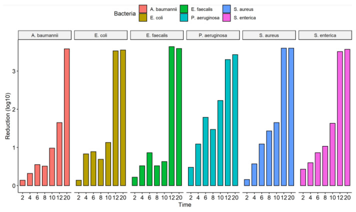
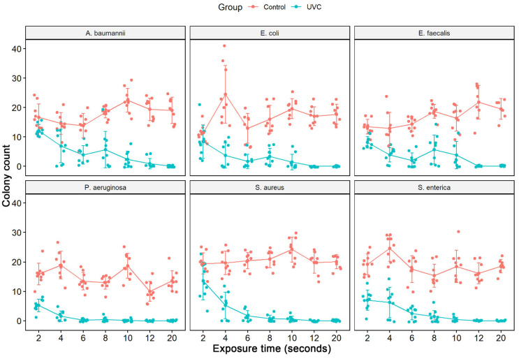
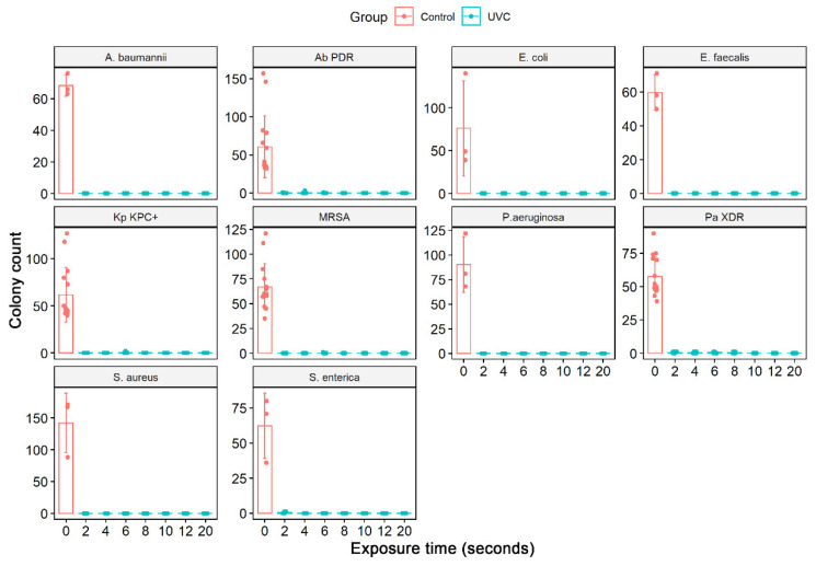
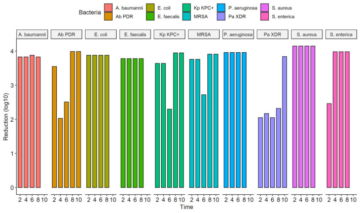
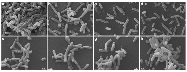
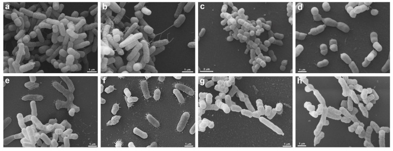
Healthcare-associated infections (HAI) worldwide includes infections by ESKAPE-E pathogens. Environmental surfaces and fomites are important components in HAI transmission dynamics, and shoe soles are vectors of HAI. Ultraviolet (UV) disinfection is an effective method to inactivate pathogenic microorganisms. In this study, we investigated whether the SANITECH UV-C shoe sole decontaminator equipment that provides germicidal UV-C radiation could effectively reduce this risk of different pathogens. Six standard strains and four clinical MDR strains in liquid and solid medium were exposed to a UV-C System at specific concentrations at other times. Bacterial inactivation (growth and cultivability) was investigated using colony counts and resazurin as metabolic indicators. SEM was performed to assess the membrane damage. Statistically significant reduction in cell viability for all ATCCs strains occurred after 10 s of exposure to the UV-C system, except for S. enterica, which only occurred at 20 s. The cell viability of P. aeruginosa (90.9%), E. faecalis and A. baumannii (85.3%), S. enterica (82.9%), E. coli (79.2%) and S. aureus (71.9%) was reduced considerably at 20 s. In colony count, after 12 s of UV-C exposure, all ATCC strains showed a 100% reduction in CFU counts, except for A. baumannii, which reduced by 97.7%. A substantial reduction of colonies above 3 log10 was observed at 12 and 20 s in all bacterial strains tested, except for A. baumannii ATCC 19606 (12 s). The exposure of ATCCs bacterial strains to the UV-C system for only 2 s was able to reduce 100% in the colony forming units (CFU) count in all ATCCs strains, S. aureus, P. aeruginosa, E. coli, A. baumannii, E. faecalis, except the S. enterica strain which had a statistically significant reduction of 99.7%. In ATCC strains, there was a substantial decrease in colonies after 4 s (sec) of exposure to the UV-C system, with a reduction ranging from 3.78-4.15 log10 CFU/mL. This reduction was observed in MDR/ESKAPE-E strains within 10 s, showing that UV-C could eliminate above 3.84 log10 CFU/mL. SEM showed a reduction of pili-like appendages after UV-C treatment in all strains except for E. coli (ATCC 25922). The Sanitech UV-C shoe sole decontaminator equipment from Astech Serv. and Fabrication Ltd. (Petrópolis, Brazil), effectively killed in vitro a series of ATCCs and MDR/ESKAPE-E bacteria of sanitary interest, commonly found in the hospital environment.

Keywords: ESKAPE-E pathogens; SEM; UV-C; cell viability; disinfection; multidrug resistance; shoe sole decontaminator.


Conflict of interest statement

The authors declare no conflict of interest.

Figures

Figure 1
Figure 1
Analysis of cell viability by the addition of resazurin after treatment with UV-C system and control group (no treatment) in different bacterial strains ATCCs (S. aureus (ATCC 6538), P. aeruginosa (ATCC 15442), S. enterica (ATCC 10708), E. coli (ATCC 25922), A. baumannii (ATCC 19606) and E. faecalis (ATCC 29212)). The measured fluorescence intensity (Relative Fluorescence Units, RFU) after the conversion of resazurin to resofurin by viable bacteria was performed either in the control group (no treatment) as in the bacterial suspensions (1:100 dilution) after exposure to the UV-C (2, 4, 6, 8, 10, 12 and 20 s). Results represent values from 3 experiments in triplicate. [times (sec) of UV-C exposure].
Figure 2
Figure 2
Efficacy of the UV-C system in reducing the number of colony-forming units of the ATCCs strains used and expressed in log10 reduction of CFU/mL; CFU, colony forming units.
Figure 3
Figure 3
Counting the number of colonies forming units (CFU/mL) of the assay from the liquid medium in different ATCCs bacterial strains (S. aureus (ATCC 6538), P. aeruginosa (ATCC 15442), S. enterica (ATCC 10708), E. coli (ATCC 25922), A. baumannii (ATCC 19606) and E. faecalis (ATCC 29212)). The number of CFU/mL was quantified in the control group (no treatment) and in the bacterial suspensions (1:100 dilution) after exposure to the UV-C system for 2, 4, 6, 8, 10, 12, and 20 s.
Figure 4
Figure 4
Quantifying the number of CFU/mL in the solid medium of the different bacterial strains ATCCs (S. aureus (ATCC 6538), P. aeruginosa (ATCC 15442), S. enterica (ATCC 10708), E. coli (ATCC 25922), A baumannii (ATCC 19606), and E. faecalis (ATCC 29212) and MDR strains representative of the ESKAPE-E group S. aureus (MRSA), P. aeruginosa (XDR), A. baumannii (PDR) and K. pneumoniae (KPC+)). In addition, the number of CFU/mL was quantified in the bacterial suspensions (104 CFU/mL) after exposure to the UV-C system for 2, 4, 6, 8, and 10 s and in the control group (no treatment). Boxes represent median and lines standard deviations.
Figure 5
Figure 5
Efficacy of the UV-C system in reducing several bacterial strains expressed in log10 reduction of CFU/mL; CFU, colony forming units; A. baumannii, Acinetobacter baumanni; Ab PDR, Acinetobacter baumannii Pandrug-resistant; E. coli, Escherichia coli; E. faecalis, Enterococcus faecalis; Kp KPC+, Klebsiella pneumoniae produtora de carbapenemase (KPC+); MRSA, Staphylococcus aureus resistente à meticilina; P. aeruginosa, Pseudomonas aeruginosa; P. aeruginosa XDR, Pseudomonas aeruginosa, Extensively drug-resistant; S. aureus, Staphylococcus aureus; S. enterica, Salmonella enterica.
Figure 6
Figure 6
Morphological analysis of UV-C treatment (2 s) by Scanning Electron Microscopy. S. enterica (ATCC 10708) (ad) and E. coli (ATCC 25922) (eh) are seen without (a,b,e,f) and under UV-C treatment (c,d,g,h). Untreated S. enterica (a,b) are elongated, presenting many pili-like appendages emerging from different regions of the cell body. Note that the cells form large aggregates, where more pili-like appendages are seen contacting each other (a). After treatment with UV-C, the S. enterica (c,d) still presents the same morphology, but there is a decrease in pili-like appendages and the formation of cell aggregates (c,d). Both, untreated (e,f) and treated (g,h) E. coli present the same morphology, the cells are elongated, and less pili-like appendages are observed (eh). There is no difference between untreated and treated E. coli.
Figure 7
Figure 7
Morphological analysis of A. baumannii (PDR) and K. pneumoniae (KPC+) by Scanning Electron Microscopy. A. baumannii (PDR) (ad) and K. pneumoniae (KPC+) (eh) are seen without (a,b,e,f) and under UV-C treatment by 2 s (c,d,g,h). Untreated A. baumannii (PDR) (a,b) presents a heterogeneous morphology, note the presence of cells with different sizes (a,b), some cells present pili-like appendages extending to other bacteria (b). After A. baumannii (PDR) treatment (c,d), the cell population is homogenously smaller (c) when compared with untreated ones (a), and no pili-like appendages are seen (d). UV-C unexposed K. pneumoniae (KPC+) (e,f) present several pili-like appendages around the elongated cell body (e,f), these projections are seen mainly when the bacteria are adhered to the substrate (f). In treated K. pneumoniae (KPC+) (g,h) it is possible to note that the cell surface is wrinkled (g,h), there are different cell sizes (g) and there are no pili-like appendages (g,h).

Similar articles

Cited by

References

    1. Tabah A., Koulenti D., Laupland K., Misset B., Valles J., de Carvalho F.B., Paiva J.A., Çakar N., Ma X., Eggimann P., et al. Characteristics and determinants of outcome of hospital-acquired bloodstream infections in intensive care units: The EUROBACT International Cohort Study. Intensiv. Care Med. 2012;38:1930–1945. doi: 10.1007/s00134-012-2695-9. - DOI - PubMed
    1. Tacconelli E., Carrara E., Savoldi A., Harbarth S., Mendelson M., Monnet D.L., Pulcini C., Kahlmeter G., Kluytmans J., Carmeli Y., et al. Discovery, research, and development of new antibiotics: The WHO priority list of antibiotic-resistant bacteria and tuberculosis. Lancet Infect. Dis. 2018;18:318–327. doi: 10.1016/S1473-3099(17)30753-3. - DOI - PubMed
    1. Li S., Eisenberg J.N.S., Spicknall I.H., Koopman J.S. Dynamics and Control of Infections Transmitted from Person to Person Through the Environment. Am. J. Epidemiol. 2009;170:257–265. doi: 10.1093/aje/kwp116. - DOI - PubMed
    1. Rashid T., VonVille H.M., Hasan I., Garey K.W. Shoe soles as a potential vector for pathogen transmission: A systematic review. J. Appl. Microbiol. 2016;121:1223–1231. doi: 10.1111/jam.13250. - DOI - PubMed
    1. Agarwal M., Hamilton-Stewart P., Dixon R.A. Contaminated operating room boots: The potential for infection. Am. J. Infect. Control. 2002;30:179–183. doi: 10.1067/mic.2002.119513. - DOI - PubMed

Publication types

LinkOut - more resources