. 2022 Dec 5;13(12):1119.
doi: 10.3390/insects13121119.

Transcriptomic Analysis Insight into the Immune Modulation during the Interaction of Ophiocordyceps sinensis and Hepialus xiaojinensis

Affiliations

Transcriptomic Analysis Insight into the Immune Modulation during the Interaction of Ophiocordyceps sinensis and Hepialus xiaojinensis

Xinxin Tong et al. Insects. .

Abstract

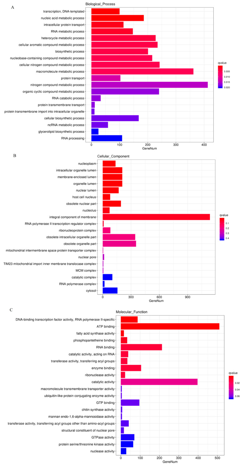
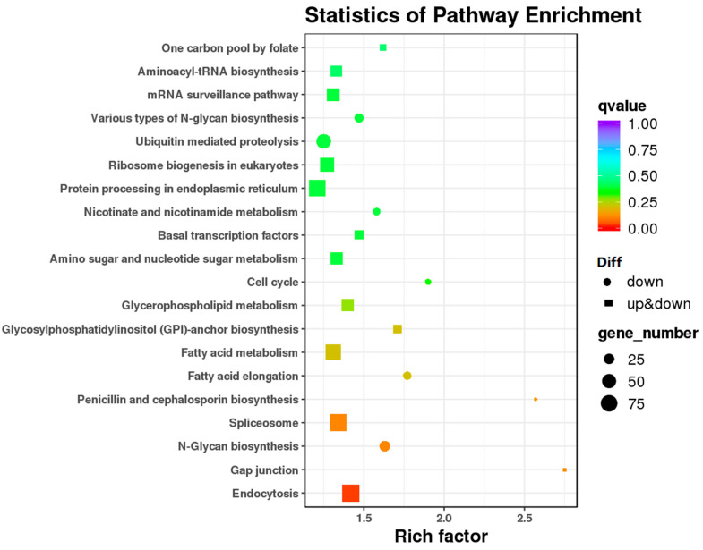
Ophiocordyceps sinensis (Berk.) is an entomopathogenic fungus that can infect the larva of the ghost moth, Hepialus xiaojinensis, causing mummification after more than one year. This prolonged infection provides a valuable model for studying the immunological interplay between an insect host and a pathogenic fungus. A comparative transcriptome analysis of pre-infection (L) and one-year post-infection (IL) larvae was performed to investigate the immune response in the host. Here, a total of 59,668 unigenes were obtained using Illumina Sequencing in IL and L. Among the 345 identified immune-related genes, 83 out of 86 immune-related differentially expressed genes (DEGs) had a much higher expression in IL than in L. Furthermore, the immune-related DEGs were classified as pathogen recognition receptors (PRRs), signal modulators or transductors, and immune effector molecules. Serpins and protease inhibitors were found to be upregulated in the late phase of infection, suppressing the host’s immune response. Based on the above analysis, the expression levels of most immune-related genes would return to the baseline with the immune response being repressed in the late phase of infection, leading to the fungal immunological tolerance after prolonged infection. Meanwhile, the transcriptomes of IL and the mummified larva (ML) were compared to explore O. sinensis invasion. A total of 1408 novel genes were identified, with 162 of them annotated with putative functions. The gene families likely implicated in O. sinensis pathogenicity have been identified, primarily including serine carboxypeptidase, peroxidase, metalloprotease peptidase, aminopeptidases, cytochrome P450, and oxidoreductase. Furthermore, quantitative real-time PCR (qPCR) was used to assess the expression levels of some critical genes that were involved in immune response and fungal pathogenicity. The results showed that their expression levels were consistent with the transcriptomes. Taken together, our findings offered a comprehensive and precise transcriptome study to understand the immune defense in H. xiaojinensis and O. sinensis invasion, which would accelerate the large-scale artificial cultivation of this medicinal fungus.

Keywords: Hepialus xiaojinensis; Ophiocordyceps sinensis; RNA-seqs; immunological interaction.


Conflict of interest statement

The authors declare no conflict of interest.

Figures

Figure 1
Figure 1
O. sinensis samples collection for transcriptome sequencing. IL represents one-year post-infected larva of H. xiaojinensis by O. sinensis. L represents the pre-infected larva of H. xiaojinensis. ML represents the mummified larva of H. xiaojinensis. Scale = 1 cm.
Figure 2
Figure 2
The analysis of differentially expressed genes (DEGs) in IL and L. The histograms of DEGs in groups. (A). The number of DEGs is shown on the top of histograms. (B). The volcano map of the DEGs in IL vs. L, with each point indicating the differentially expressed genes and the abscissa representing the logarithm of the expression multiple of a gene in the two groups. Green represents downregulation of gene expression in L compared to IL, red represents upregulation of gene expression in L compared to IL, and black dots represent genes with no significant expression differences. (C). Hierarchical clustering of differentially expressed genes. Each column represents one sample. Rows represent different genes. The expression level of genes (FPKM) was normalized by log2, and blue color represents the downregulated gene, while red color represents the upregulated genes. L represents the pre-infected larva of H. xiaojinensis. IL represents the one year post-infected larva of H. xiaojinensis by O. sinensis.
Figure 3
Figure 3
The most enriched GO functional classification of DEGs in IL vs. L. The most enriched GO terms in Biological_Process (A), Cellular_Component (B) and Molecular_Function (C) were presented. X-axis represents the gene number of top GO terms enriched among DEGs. L represents the pre-infected larva of H. xiaojinensis. IL represents the one-year post-infection larva of H. xiaojinensis by O. sinensis.
Figure 4
Figure 4
The scatter diagram of the enriched KEGG pathway of DEGs in IL vs. L. Y-axis: pathway; X-axis: Enrichment factor. The color of the dots stands for q-value. ‘Diff’’ represents differential expression. Circle represents the downregulated DEGs in this pathway, and rectangle represents the upregulated and downregulated DEGs in this pathway. The size of the dots represents the number of DEGs enriched in this pathway. IL represents the one-year post-infected larva of H. xiaojinensis by O. sinensis. L represents the pre-infected larva of H. xiaojinensis.
Figure 5
Figure 5
The heatmap of the cluster diagram of differentially expressed immune-related gene expression patterns in IL vs. L. Each column represents one sample. Rows represent different genes. The expression level of genes (FPKM) was normalized by log2 and presented as different colors based on scale bar. L represents the pre-infected larva of H. xiaojinensis. IL represents the one-year post-infected larva of H. xiaojinensis by O. sinensis.
Figure 6
Figure 6
Schematic structure of H. xiaojinensis PGPRs and βGRPs identified in the IL and L. The sequence of βGRP 1-3 in H. xiaojinensis is truncated at the N-terminus. The putative signal peptide (SP), otransmembrane domain (TR), PGRP homologous domain (PGRP), β-1,3-glucan binding domain (CM39), and β-glucanase-like domain (CH16) are indicated in different colored boxes. Lengths of the amino acid sequences are indicated. PGRP-L represents long-type PGRP. PGRP-S represents short-type PGRP. L represents the pre-infected larva of H. xiaojinensis. IL represents the one-year post-infected larva of H. xiaojinensis by O. sinensis.
Figure 7
Figure 7
(A) Schematic structure of H. xiaojinensis CSP_14D. (B) Crystal structure of H. xiaojinensis CSP (BMK_Unigene_108359) predicted by AlphaFold 2. (C) Multiple alignments of the amino acid sequence of CSP with homologs from other insect species. Grey ‘*’ represents 100% identity, grey ‘:’ represents ≥ 70% identity, grey ‘.’ represents 50% identity. Clip domains and serine protease domains are in orange and blue frames, respectively. Green strip represents signal peptide region. Purple strip represents linker region. The conserved cysteine residues responsible for disulfide bonds are labeled with red triangles. CSP: Clip domain containing serine protease.
Figure 8
Figure 8
The putative pathways, on the basis of Drosophila research, mediate the response to pathogen infection. The component of the fungal cell wall is sensed by circulating βGRP, peptidoglycan recognition proteins (PGRPs, e.g., PGRP-SA, PGRP-SD and PGRP-LB), a process that activates a proteinase-signaling cascade followed by cleavage of the proSpz (ProSpaetzle) to mature Spz (Spaetzle), binds with Toll, which causes aggregation of intracellular proteins, e.g., MyD88, leading to expression of anti-microbiology peptide (AMP) genes; with the response to the pathogen components, C type lectin (CLT) and/or scavenger receptors (SRs type C) activates JAK/STAT signaling pathway, leading to the expression of pro-apoptotic genes; PGRP-LE in hemolymph activates the prophenoloxidase (proPO) cascade followed by cleavage of the proPO to mature phenoloxidase PO, leading to the melanization for fungi infection; PGRP-LE in cytoplasm activates autophagy-mediated immunity defense; with the responses to fungal infection, PGRP-LC activates Imd pathway, leading to the expression of AMP genes. ‘-’ indicates the name of pathway, the red triangles and letters represent ‘upregulated’ proteins in IL compared to L; the blue triangle and letters represent the ‘downregulated’ protein in IL compared to L.
Figure 9
Figure 9
(A). The histograms of DEGs in groups; the number of DEGs is shown on the top of histograms. (B). The volcano map of DEGs in IL vs. ML; each point represents the differentially expressed gene and the abscissa represents the logarithm of the expression multiple of a gene in the two groups. Green and red dots represent genes with significant expression differences, green represents downregulation of gene expression in ML compared to IL, red represents upregulation of gene expression in ML compared to IL, and black dots represent genes with no significant expression differences. IL represents the one − year post − infection larva of H. xiaojinensis by O. sinensis. ML represents the mummified larva of H. xiaojinensis.
Figure 10
Figure 10
(AC) The map of the GO classification of DEGs in IL vs. ML. X-axis is the percentage of the genes annotated to this term in total number of annotated genes. The Y-axis stands for GO term. IL represents the one-year post-infected larva of H. xiaojinensis by O. sinensis. ML represents the mummified larva of H. xiaojinensis.
Figure 11
Figure 11
The Scatter diagram of the enriched KEGG pathway of DEGs in IL vs. ML. axis. Pathway; X-axis: Enrichment factor. Enrichment factor is calculated as ratio of DEGs annotated to the term over all DEGs. The color of the dots stands for q-value. ‘Diff’ represents differential expression. Rectangle represents the upregulated and downregulated DEGs in this pathway. IL represents the one-year post-infected larva of H. xiaojinensis by O. sinensis. ML represents the mummified larva of H. xiaojinensis.
Figure 12
Figure 12
The heatmap of the Cluster diagram of DGEs probably involved in fungal pathogenicity in IL vs. ML. Each column represents one sample. Rows represent different genes. The expression level of genes (FPKM) was normalized by log2 and presents as different colors based on scale bar. IL represents the one-year post-infected larva of H. xiaojinensis by O. sinensis. ML represents the mummified larva of H. xiaojinensis.
Figure 13
Figure 13
qPCR analysis using gene − specific primers was performed with cDNA samples of L, IL and ML. H. xiaojinensis larvae were O. sinensis-challenged and collected in one year after the infection, as indicated. (A,B): Log2 fold change comparison with standard deviation, Elongation factor protein gene was used as an internal control to normalize the amount of template in IL vs. L in A. 18s rRNA was used as an internal control to normalize the amount of template in IL vs. ML in (C,D): correlation analysis between RNA-seq and qPCR log 2 (fold-change) results from the same RNA samples (p value = 0.022 and 0.0266 in IL vs. L and IL vs. MF, respectively). Dots represent relative expression of genes, lines represent the regression lines, dash lines represent 95% confidence interval. These experiments were implemented at least in triplicate. L represents the pre-infected larva of H. xiaojinensis. IL represents the one-year post-infected larva of H. xiaojinensis by O. sinensis. ML represents the mummified larva of H. xiaojinensis.

References

    1. Wei Y., Zhang L., Wang J., Wang W., Niyati N., Guo Y., Wang X. Chinese caterpillar fungus (Ophiocordyceps sinensis) in China: Current distribution, trading, and futures under climate change and overexploitation. Sci. Total Environ. 2021;755:142548. doi: 10.1016/j.scitotenv.2020.142548. - DOI - PMC - PubMed
    1. Zhang Y.J., Li E., Wang C.S., Li Y.L., Liu X.Z. Ophiocordyceps sinensis, the flagship fungus of China: Terminology, life strategy and ecology. Mycology. 2012;3:2–10.
    1. Xu J., Huang Y., Chen X.X., Zheng S.C., Chen P., Mo M.H. The Mechanisms of Pharmacological Activities of Ophiocordyceps sinensis Fungi. Phytother. Res. 2016;30:1572–1583. doi: 10.1002/ptr.5673. - DOI - PubMed
    1. Guo J.L., Liu X.Y., Kanari K. Economic aspects of harvesting and trading the Chinese Caterpillar Fungus Ophiocordyceps sinensis and Southern Schisandra Schisandra sphenanthera in China’s Upper Yangtze Ecoregion. Traffic Bull. 2012;24:15–24.
    1. Li X., Liu Q., Li W., Li Q., Qian Z., Liu X., Dong C. A breakthrough in the artificial cultivation of Chinese cordyceps on a large-scale and its impact on science, the economy, and industry. Crit. Rev. Biotechnol. 2019;39:181–191. doi: 10.1080/07388551.2018.1531820. - DOI - PubMed

LinkOut - more resources