Review
. 2022 Dec 7;23(24):15479.
doi: 10.3390/ijms232415479.

The Role of microRNAs in Inflammation

Affiliations
Review

The Role of microRNAs in Inflammation

Kaushik Das et al. Int J Mol Sci. .

Abstract

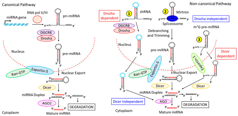
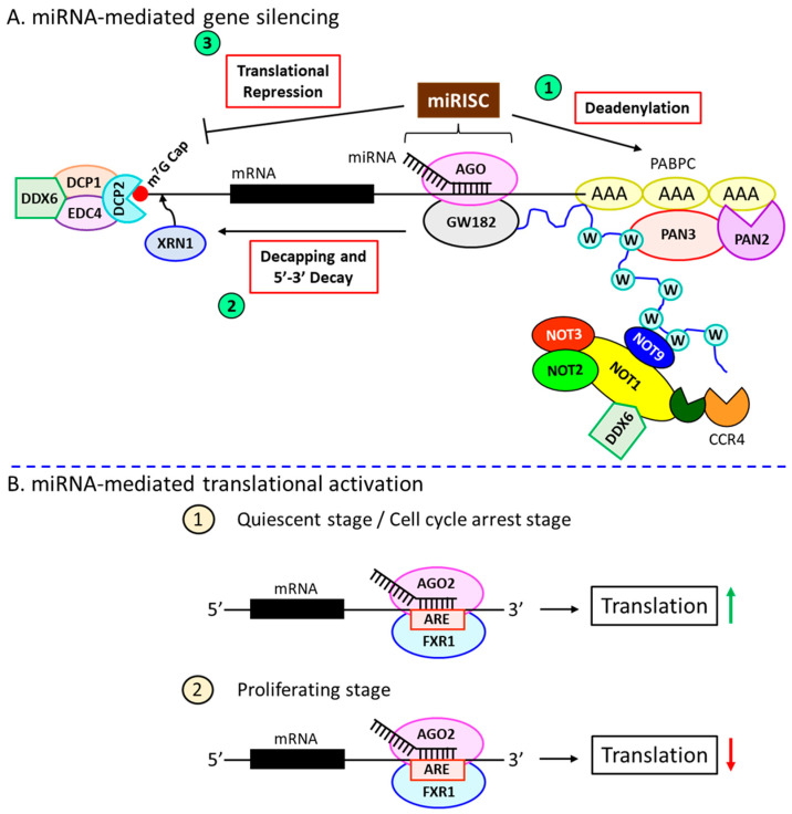
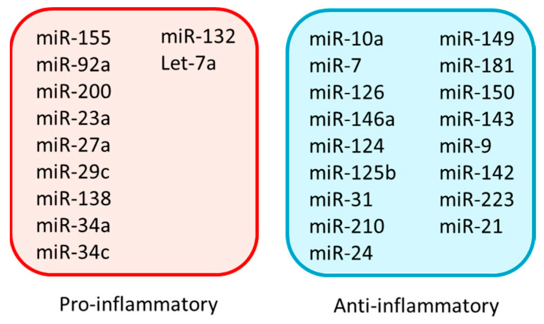
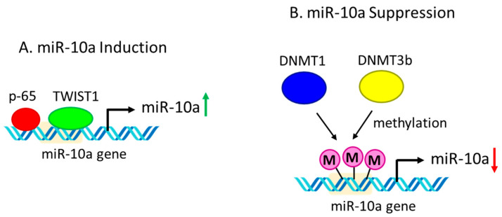
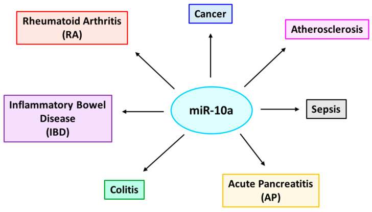
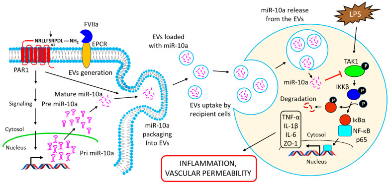
Inflammation is a biological response of the immune system to various insults, such as pathogens, toxic compounds, damaged cells, and radiation. The complex network of pro- and anti-inflammatory factors and their direction towards inflammation often leads to the development and progression of various inflammation-associated diseases. The role of small non-coding RNAs (small ncRNAs) in inflammation has gained much attention in the past two decades for their regulation of inflammatory gene expression at multiple levels and their potential to serve as biomarkers and therapeutic targets in various diseases. One group of small ncRNAs, microRNAs (miRNAs), has become a key regulator in various inflammatory disease conditions. Their fine-tuning of target gene regulation often turns out to be an important factor in controlling aberrant inflammatory reactions in the system. This review summarizes the biogenesis of miRNA and the mechanisms of miRNA-mediated gene regulation. The review also briefly discusses various pro- and anti-inflammatory miRNAs, their targets and functions, and provides a detailed discussion on the role of miR-10a in inflammation.

Keywords: inflammation; microRNA; small non-coding RNA.


Conflict of interest statement

The authors declare no conflict of interest.

Figures

Figure 1
Figure 1
Biogenesis pathways of miRNAs. (Left panel) Canonical pathway. miRNA genes are transcribed in the nucleus by RNA Pol-II or -III to generate pri-miRNAs, which are processed by DGCR8-Drosha to produce pre-miRNAs. pre-miRNAs are transported via Exportin-5-Ran-GTP into the cytoplasm, followed by further processing by Dicer to generate the miRNA duplex. miRNA duplex then binds to AGO2, which further processes the duplex to produce one mature miRNA strand while degrading the other strand. (Right panel) Non-canonical pathway. The non-canonical pathway consists of multiple pathways: ① In Drosha-dependent but Dicer-independent pathway, shRNAs are processed by DGCR8-Drosha, and are readily transported from the nucleus to the cytoplasm by Exportin-5-Ran-GTP. The transported miRNA duplex is processed by AGO2 (designated as AGO in the figure) to generate the mature miRNA. ② In one Drosha-independent and Dicer-dependent pathway, Mirtrons, associated with spliceosomes, undergo debranching and trimming to generate pre-miRNAs, which are transported into the cytoplasm by Exportin-5-Ran-GTP, followed by sequential processing by Dicer and AGO1-4, respectively, to produce mature miRNAs. ③ In another Drosha-independent and Dicer-dependent pathway, (m7G)-capped pre-miRNAs are released into the cytoplasm via exportin-1, followed by Dicer processing to form miRNA duplex, which is further processed by AGO1-4 to generate mature miRNAs.
Figure 2
Figure 2
miRNAs-mediated gene regulation. (A) miRNA-mediated gene silencing mechanisms. miRNA-dependent gene silencing occurs in three independent pathways. miRNA-bound AGO forms the miRISC complex, which plays a central role in gene regulation. ① miRISC recruits the scaffolding protein, GW182, which further recruits effector complexes PAN2-3 and CCR4-NOT to induce the deadenylation process. Deadenylation is enhanced by the interaction among GW182, PABPC, and CCR4-NOT through tryptophan (W) repeats. ② GW182 recruits the decapping protein, DCP2, to the 5’-m7G cap of the target mRNA molecules along with other associate proteins (such as EDP4, DCP1, and DDX6), resulting in the decapping at the 5’ terminus. The decapped 5’-terminus becomes vulnerable to cleavage by XRN1 due to its 5’-3’ exonuclease activity. ③ miRISC-GW182 also interferes with the binding of ribosomes at the 5’ end of the mRNA molecules, resulting in translational repression. (B) miRNA-mediated translational activation. ① AGO2 and FXR1 bind to the AREs at the 3’-UTR of the target mRNAs in the quiescent stage or during cell cycle arrest and induce the translational process. ② The same binding in proliferating stage suppresses the translation.
Figure 3
Figure 3
A list of pro- and anti-inflammatory miRNAs in the context of various disease conditions.
Figure 4
Figure 4
Regulation of miR-10a expression. (A) miR-10a induction. Transcription factors p65 and TWIST1 bind to the promoter region of the miR-10a gene resulting in the induction of miR-10a expression. (B) miR-10a suppression. DNA methyltransferases DNMT1 and DNMT3b cause promoter methylation of the miR-10a gene resulting in the down-regulation of miR-10a expression.
Figure 5
Figure 5
Various inflammation-associated diseases regulated by miR-10a. miR-10a is a key regulatory molecule that influences several inflammation-associated diseases, such as rheumatoid arthritis (RA), inflammatory bowel disease (IBD), colitis, acute pancreatitis (AP), sepsis, atherosclerosis, and cancer.
Figure 6
Figure 6
Schematic representation showing the release of miR-10a-loaded EVs from the endothelial cells and their uptake by target recipient cells to alter their phenotypic responses. FVIIa binds to EPCR on endothelial cells and activates PAR1 by proteolytic cleavage at the R41 site. The activation of PAR1 leads to the induction of miR-10a expression in the cells. PAR1 activation also triggers the release of EVs from the endothelial cells, and the FVIIa-EEVs are packaged with miR-10a. The EEVs are taken up by the target recipient cells (such as monocytes or naïve endothelial cells) via endocytosis, and the release of miR-10a from the vesicles into the recipient cell’s cytosol results in the down-regulation of the target gene’s TAK1 expression. TAK1 down-regulation impairs LPS-induced activation of the NF-κB signaling pathway and the concomitant induction of pro-inflammatory cytokines, TNF-α, IL-1β, and IL-6, as well as tight junction protein ZO-1, thereby limiting LPS-induced inflammation and vascular permeability in the recipient cells.

References

    1. Cronkite D.A., Strutt T.M. The Regulation of Inflammation by Innate and Adaptive Lymphocytes. J. Immunol. Res. 2018;2018:1467538. doi: 10.1155/2018/1467538. - DOI - PMC - PubMed
    1. Parke D.V., Parke A.L. Chemical-induced inflammation and inflammatory diseases. Int. J. Occup. Med. Environ. Health. 1996;9:211–217. - PubMed
    1. Medzhitov R. Inflammation 2010: New adventures of an old flame. Cell. 2010;140:771–776. doi: 10.1016/j.cell.2010.03.006. - DOI - PubMed
    1. Schaue D., Micewicz E.D., Ratikan J.A., Xie M.W., Cheng G., McBride W.H. Radiation and inflammation. Semin. Radiat. Oncol. 2015;25:4–10. doi: 10.1016/j.semradonc.2014.07.007. - DOI - PMC - PubMed
    1. Ferrero-Miliani L., Nielsen O.H., Andersen P.S., Girardin S.E. Chronic inflammation: Importance of NOD2 and NALP3 in interleukin-1beta generation. Clin. Exp. Immunol. 2007;147:227–235. doi: 10.1111/j.1365-2249.2006.03261.x. - DOI - PMC - PubMed

LinkOut - more resources