Review
. 2022 Dec 7;23(24):15489.
doi: 10.3390/ijms232415489.

Non-Alcoholic Fatty Liver Disease (NAFLD) Pathogenesis and Natural Products for Prevention and Treatment

Affiliations
Review

Non-Alcoholic Fatty Liver Disease (NAFLD) Pathogenesis and Natural Products for Prevention and Treatment

Xiangyu Guo et al. Int J Mol Sci. .

Abstract

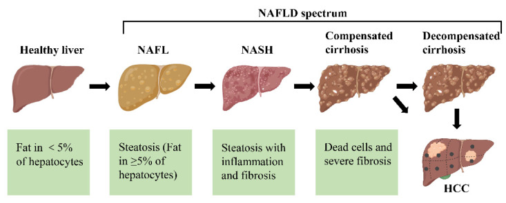
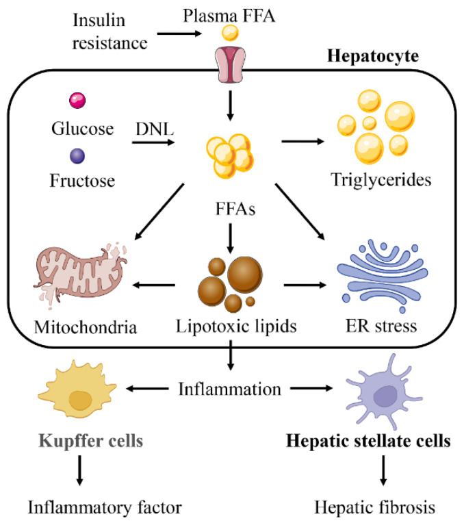
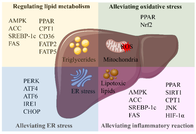
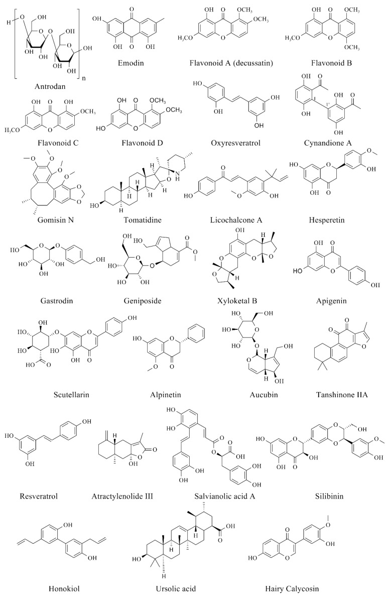
Non-alcoholic fatty liver disease (NAFLD) is the most prevalent chronic liver disease, affecting approximately one-quarter of the global population, and has become a world public health issue. NAFLD is a clinicopathological syndrome characterized by hepatic steatosis, excluding ethanol and other definite liver damage factors. Recent studies have shown that the development of NAFLD is associated with lipid accumulation, oxidative stress, endoplasmic reticulum stress, and lipotoxicity. A range of natural products have been reported as regulators of NAFLD in vivo and in vitro. This paper reviews the pathogenesis of NAFLD and some natural products that have been shown to have therapeutic effects on NAFLD. Our work shows that natural products can be a potential therapeutic option for NAFLD.

Keywords: NAFLD; endoplasmic reticulum stress; lipid accumulation; lipotoxicity; natural products; oxidative stress; pathogenesis; plants.


Conflict of interest statement

The authors declare no conflict of interest.

Figures

Figure 1
Figure 1
The different stages of non-alcoholic fatty liver disease (NAFLD). First, the healthy livers develop non-alcoholic fatty liver (NAFL) with hepatocellular steatosis as the main feature. If left untreated, NAFL may progress to a more severe form of non-alcoholic steatohepatitis (NASH), defined as inflammation and fibrosis in addition to hepatocellular steatosis. As the disease progresses, NASH may progress to cirrhosis and even to hepatocellular carcinoma (HCC).
Figure 2
Figure 2
Abnormal lipid accumulation in NAFLD. The increase in hepatic lipid accumulation is due to the absorption of large amounts of free fatty acids (FFAs) synthesized triglycerides by the liver from white adipose tissue (WAT), high-fat and high-sugar foods, and de novo lipogenesis (DNL). Insulin resistance plays a vital role in this process. Insulin resistance promotes glucose absorption and enhances the lipolysis of WAT. This leads to the activation of the DNL pathway. Abbreviations: ChREBP, carbohydrate response element binding protein; SREBP-1c, sterol regulatory element binding protein 1c; ACC, acetyl-CoA carboxylase; FAS, fatty acid synthase.
Figure 3
Figure 3
Fatty acid oxidation system in NAFLD. The fatty acid oxidation system consists of peroxisome, mitochondria and microsomes. Mitochondria play a vital role in fatty acid oxidation and energy supply. Glucose enhanced glycolysis and increased pyruvate content through the de novo lipogenesis (DNL) pathway. Pyruvate enters mitochondria and is converted into acetyl-CoA. Part of acetyl-CoA enters tricarboxylic acid cycle (TCA) and then synthesizes free fatty acids (FFAs) through the DNL pathway. The synthesized FFAs enter mitochondria together with the plasma FFAs through carnitine palmitoyltransferase 1 (CPT1) and are converted into acyl-CoA. Acyl-CoA is converted into acetyl-CoA by β-oxidation and enters the TCA to generate energy. The components of the mitochondrial respiratory chain are abnormally reduced by electrons and react with oxygen, producing a large number of reactive oxygen species (ROS). ROS further oxidize lipid deposition to form lipid peroxide, which leads to inflammatory reaction. Abbreviations: ETC, electron transport chain; PPAR, peroxisome proliferation-activated receptor.
Figure 4
Figure 4
Endoplasmic reticulum (ER) stress in NAFLD. With the increase of lipid accumulation, ER stress leads to a large number of unfolded proteins, thus triggering unfolded protein response (UPR). UPR is mediated by protein kinase RNA-like ER kinase (PERK), inositol-requiring enzyme 1 (IRE1), and activating transcription factor 6 (ATF6). PERK-mediated phosphorylation of eukaryotic initiation factor 2α (eIF2α) leads to the transient weakening of translation, but activation of transcription factor 4 (ATF4) induces the gene expression of CCAAT-enhancer-binding protein homologous protein (CHOP). ATF6 can also activate CHOP to induce apoptosis. ATF6 and IRE1 promote the expression of X-box binding protein-1 (XBP1), and mediate inflammation through the c-Jun N-terminal kinase (JNK) signaling pathway. IRE1 can also directly promote the activation of JNK and activate tumor necrosis factor (TNF) receptor-related factor 2 (TRAF2), thus promoting cell apoptosis. Abbreviations: GRP78, glucose-regulated protein 78.
Figure 5
Figure 5
Lipotoxicity is the core influencing factor for NAFLD to develop into a more serious situation. Lipotoxicity will aggravate mitochondrial dysfunction and endoplasmic reticulum (ER) stress caused by free fatty acids (FFAs). Lipotoxicity can also induce Kupffer cells and hepatic stellate cells to produce a wider range of inflammatory reactions and liver fibrosis.
Figure 6
Figure 6
Related targets of natural products in the treatment of NAFLD. Abbreviations: AMPK, (AMP)-activated protein kinase; ACC, acetyl-CoA carboxylase; SREBP-1c, sterol regulatory element-binding protein 1c; FAS, fatty acid synthase; PPAR, peroxisome proliferation-activated receptor; CPT1, carnitine palmitoyltransferase 1; CD36, cluster of differentiation 36; FATP2, fatty acid transport proteins 2; FATP5, fatty acid transport proteins 5; Nrf2, nuclear factor erythroid-derived 2-like 2; PERK, protein kinase RNA-like ER kinase; ATF4, activating transcription factor 4; ATF6, activating transcription factor 6; IRE1, inositol-requiring enzyme 1; CHOP, CCAAT-enhancer-binding protein homologous protein; SIRT1, silent information regulator 1; JNK, c-Jun N-terminal kinase; HIF-1α, hypoxia-inducible factor 1α.
Figure 7
Figure 7
Chemical structures of bioactive compounds for relieving NAFLD.

References

    1. Chalasani N., Younossi Z., Lavine J.E., Diehl A.M., Brunt E.M., Cusi K., Charlton M., Sanyal A.J. The diagnosis and management of non-alcoholic fatty liver disease: Practice guideline by the American Association for the Study of Liver Diseases, American College of Gastroenterology, and the American Gastroenterological Association. Hepatology. 2012;55:2005–2023. doi: 10.1002/hep.25762. - DOI - PubMed
    1. Milic S., Lulic D., Stimac D. Non-alcoholic fatty liver disease and obesity: Biochemical, metabolic and clinical presentations. World J. Gastroenterol. 2014;20:9330–9337. - PMC - PubMed
    1. Leoni S., Tovoli F., Napoli L., Serio I., Ferri S., Bolondi L. Current guidelines for the management of non-alcoholic fatty liver disease: A systematic review with comparative analysis. World J. Gastroenterol. 2018;24:3361–3373. doi: 10.3748/wjg.v24.i30.3361. - DOI - PMC - PubMed
    1. Bessone F., Razori M.V., Roma M.G. Molecular pathways of nonalcoholic fatty liver disease development and progression. Cell. Mol. Life Sci. 2019;76:99–128. doi: 10.1007/s00018-018-2947-0. - DOI - PMC - PubMed
    1. Machado M.V., Diehl A.M. Pathogenesis of nonalcoholic steatohepatitis. Gastroenterology. 2016;150:1769–1777. doi: 10.1053/j.gastro.2016.02.066. - DOI - PMC - PubMed

MeSH terms