. 2022 Dec 7;23(24):15498.
doi: 10.3390/ijms232415498.

Impacts of Telomeric Length, Chronic Hypoxia, Senescence, and Senescence-Associated Secretory Phenotype on the Development of Thoracic Aortic Aneurysm

Affiliations

Impacts of Telomeric Length, Chronic Hypoxia, Senescence, and Senescence-Associated Secretory Phenotype on the Development of Thoracic Aortic Aneurysm

Thomas Aschacher et al. Int J Mol Sci. .

Abstract

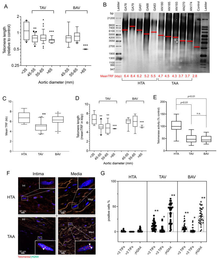
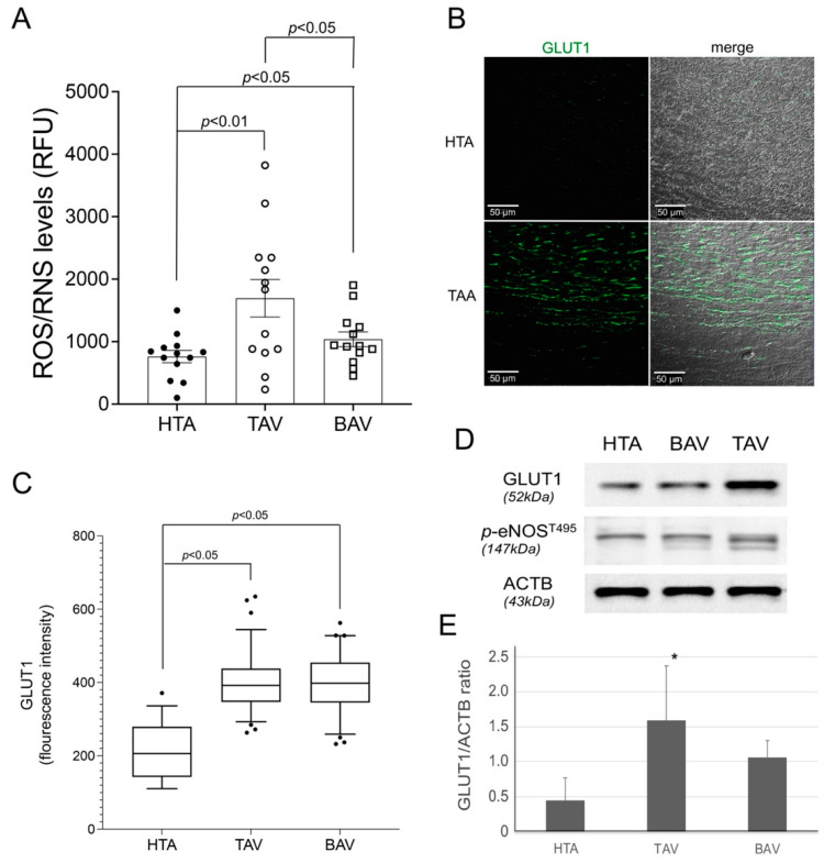
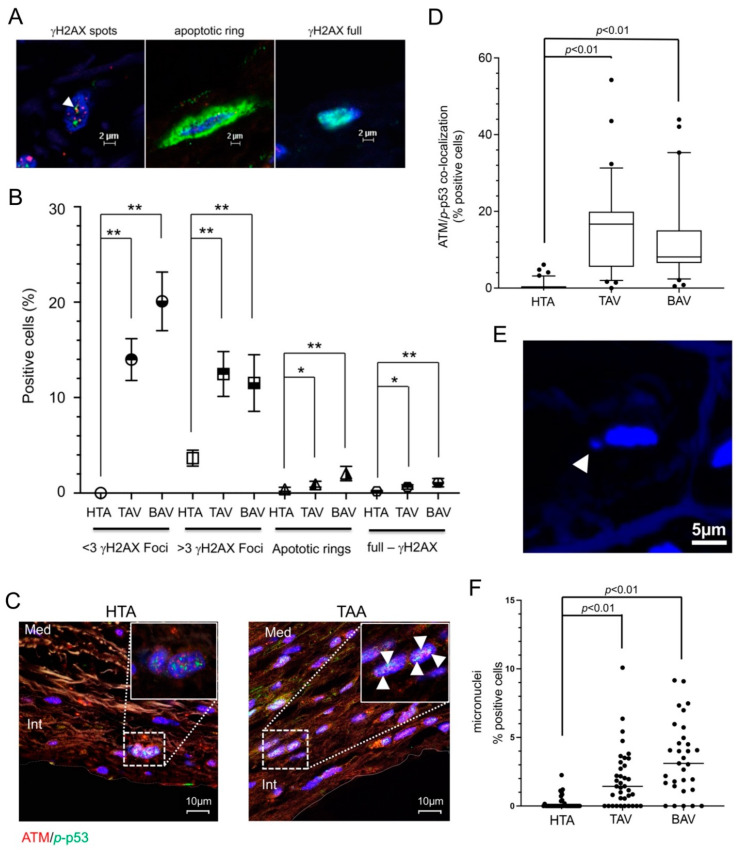
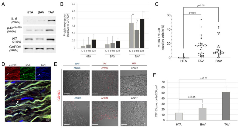
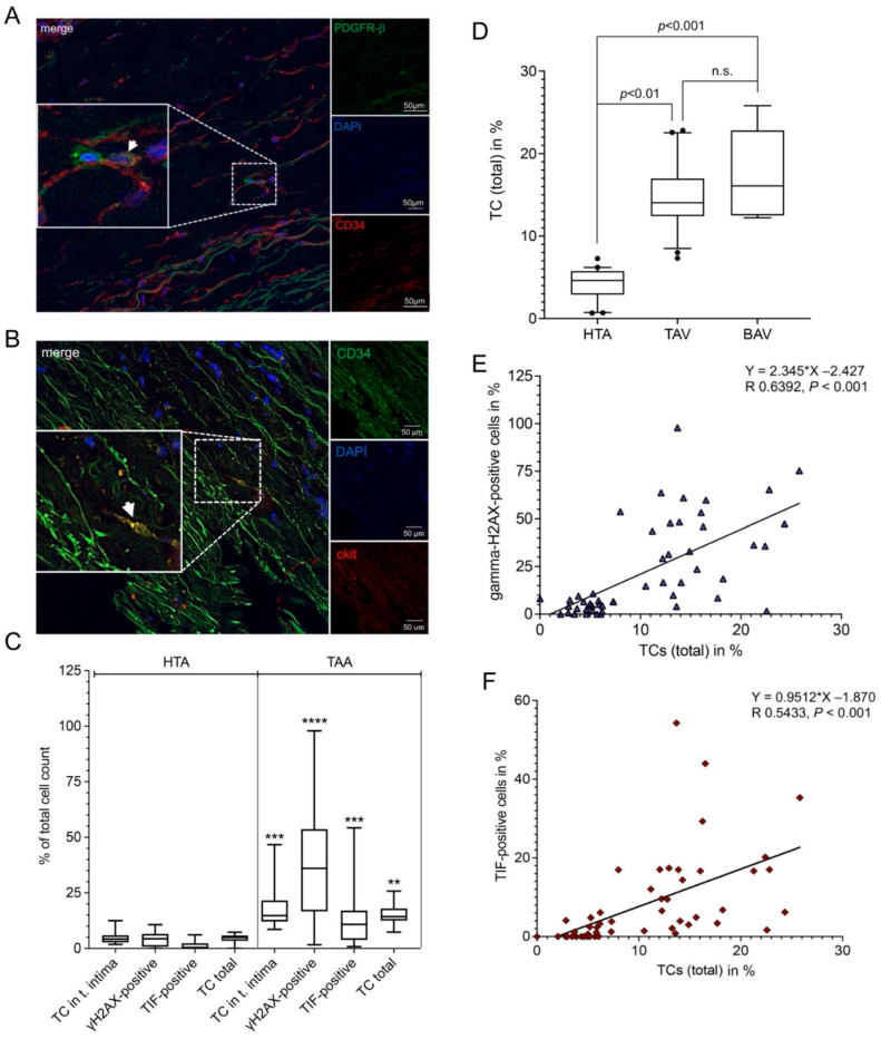
Thoracic aortic aneurysm (TAA) is an age-related and life-threatening vascular disease. Telomere shortening is a predictor of age-related diseases, and its progression is associated with premature vascular disease. The aim of the present work was to investigate the impacts of chronic hypoxia and telomeric DNA damage on cellular homeostasis and vascular degeneration of TAA. We analyzed healthy and aortic aneurysm specimens (215 samples) for telomere length (TL), chronic DNA damage, and resulting changes in cellular homeostasis, focusing on senescence and apoptosis. Compared with healthy thoracic aorta (HTA), patients with tricuspid aortic valve (TAV) showed telomere shortening with increasing TAA size, in contrast to genetically predisposed bicuspid aortic valve (BAV). In addition, TL was associated with chronic hypoxia and telomeric DNA damage and with the induction of senescence-associated secretory phenotype (SASP). TAA-TAV specimens showed a significant difference in SASP-marker expression of IL-6, NF-κB, mTOR, and cell-cycle regulators (γH2AX, Rb, p53, p21), compared to HTA and TAA-BAV. Furthermore, we observed an increase in CD163+ macrophages and a correlation between hypoxic DNA damage and the number of aortic telocytes. We conclude that chronic hypoxia is associated with telomeric DNA damage and the induction of SASP in a diseased aortic wall, promising a new therapeutic target.

Keywords: DNA damage; aneurysm; cell death; senescence-associated secretory phenotype; telocytes; telomere; thoracic aorta.


Conflict of interest statement

The authors confirm that there is no conflict of interest.

Figures

Figure 1
Figure 1
Increase in telomere damage and shortened telomeres in thoracic aortic aneurysm. Measurement of telomere length (TL) in healthy and diseased aortic tissues showed a significant shortening of telomeres and an increase in telomere-specific DNA damage. (AD) Measurement of telomeres was performed by relative qPCR method and quantitative Southern blot method. (A) Statistical analysis of qPCR measurement from the two groups TAV (tricuspid aortic valve (n = 109)) and BAV (bicuspid aortic valve, (n = 65)) compared to healthy aortic specimens (<35 mm, (n = 41)). The x-axis shows the individual sizes of the aortic tissues at the time of surgery. Specific analysis of TL in kilobase pairs (kbp) was carried out using Southern blot analysis (HTA samples, n = 16, TAA samples, n = 44). (B) shows a representative blot of the measurements. HTA, human thoracic aorta; TAA, thoracic aortic aneurysm. Mean TRF is shown in kbp along the x-axis and marked as a red line in the blot. Molecular ladder in base pairs (bp) are shown to the left and right of the blot. (C) Statistical analysis of the telomeric Southern blot evaluation for the control group HTA (n = 16) and the two TAA groups, TAV (n = 24) and BAV (n = 20), separately. Subfigure (D) shows the statistical analysis of the quantitative TL divided into the three groups HTA, TAV, and BAV, and along the x-axis divided according to the size of the aorta. (E) Relative telomerase activity measured in isolated proteins of all human samples by telomerase TRAP-ELISA assay (HTA group (n = 41) and the two TAA groups, TAV (n = 109) and BAV (n = 65)). n.s., non-significant. The measurement of telomere damage was performed by immunohistochemistry. (F) Representative images of the staining (red, telomeres; green, γH2AX) in the different aortic layers (intima and media). Cell nuclei were stained by DAPI (blue). Co-localizations, telomere-induced foci (TIFs), occurred more frequently in the TAA group vs. the HTA group. (G) Statistical analysis of TIFs divided into <3 TIFs, >3 TIFs, and γH2AX alone, plotted separately for 41 HTA samples (5707 analyzed cells), TAV samples (19,421 analyzed cells), and 65 BAV samples (13,530 analyzed cells). * p < 0.05; ** p < 0.01; *** p < 0.001.
Figure 2
Figure 2
ROS/RNS and GLUT1 expression was detected in aneurysm specimens. Measurement of ROS/RNS and GLUT1 protein levels indicated activation during hypoxic stress in aneurysm sections. (A) ROS/RNS measurement by OxiSelect™ in protein extracts of healthy aortic (HTA, n = 13) and aneurysm (TAV, n = 13, BAV, n = 13) specimens. RFU, relative fluorescence units. GLUT1 expression was measured in all aortic samples by immunofluorescence (IF, (B,C)) and confirmed in selected samples (13 samples of each group) by Western blot (D,E). (B) Representative image of GLUT1 protein (green) expression stained by IF. Scale bar, 50 µm. (C) Statistical analysis for GLUT1 protein expression in HTA (4092 cells), compared to TAA-TAV (18,293 cells) and TAA-BAV (11,543 cells) specimens. (D) The blot shows representative GLUT1 and p-eNOS specific results, and the reference beta-actin (ACTB). (E) Statistical analysis for GLUT1 protein expression detected by Western blot is given (HTA, n = 13; TAV, n = 13; BAV, n = 13). * p < 0.05.
Figure 3
Figure 3
DNA damage, cell senescence and cell death measured by γH2AX/ATM/p-p53 staining. Hypoxic stress in aneurysm sections leads to DNA damage and cellular senescence measured by γH2AX IF-staining. (A) Representative images of γH2AX spots (white arrow, left image), apoptotic ring (middle image) and γH2AX full staining (right image) detected in aortic and aneurysmal specimens. Green, γH2AX; blue, cell nuclei (DAPI). Scale bar, 2 µm. (B) Statistical calculations of different amounts of γH2AX foci (<3 foci and >3 foci), apoptotic rings, and full-γH2AX staining measured in HTA (n = 41) and both TAA groups, TAV (n = 109) and BAV (n = 65). n of counted cells: HTA, 5707; TAV, 19,421 cells; and BAV, 13,530 cells. * p < 0.05; ** p < 0.01. ATM and phosphorylated (p)-p53 expression was measured by IF staining (C,D). (C) Representative images of ATM protein (red) and p-p53 (green) expression stained by IF. Colocalizations were found in TAA samples (white arrows). Blue, cell nuclei (DAPI). Med, media; Int, intima. Scale bar, 10 µm. (D) Statistical analysis for ATM and p-p53 protein colocalization in HTA, compared to TAA-TAV and TAA-BAV specimens. ATM/p53 staining measured in HTA samples (n = 23) and both TAA groups, TAV samples (n = 61) and BAV samples (n = 53). Numbers of counted cells of each group: HTA, 3922 cells; TAV, 11,568 cells; and BAV, 9437 cells. (E) The image shows representative micronuclei next to cell nuclei stained by DAPI (blue). (F) Statistical analysis for positive cells with micronuclei (%) detected by IF is given.
Figure 4
Figure 4
Senescence and SASP expression in healthy and diseased thoracic aortic tissue. Expression of cellular marker, a chemokine (IL-6), cell cycle regulatory proteins (Rb, p21), and SASP regulatory proteins (mTOR and NF-κB). (A) Representative blot of IL-6, p-Rb, and p21 measured by Western blot, GAPDH was used as an internal control. Protein size is given in kilodaltons (kDa). HTA, human thoracic aorta; TAV, tricuspid aortic valve; BAV, bicuspid aortic valve. (B) Statistical analysis for protein expression detected by Western blot in HTA (n = 13), compared to TAA-TAV (n = 13) and TAA-BAV (n = 13) specimens. Protein expression was calculated relative to internal control (GAPDH). * p < 0.05; ** p < 0.01. NF-κB/p-mTOR protein expression given as colocalization (white arrow) was detected by IF-staining. (C) Statistical calculations for HTA (n = 24), TAV (n = 37), and BAV (n = 30)). A representative image of IF staining is shown (D). Red, p-mTOR; green, NF-κB; blue, cell nuclei (DAPI). (E) Representative image of CD163+ macrophages detected in aortic and aneurysmal specimens. Scale bar, 100 µm. (F) Statistical calculations of CD163-positive cells/200 µm2 in HTA, TAV, and BAV specimens.
Figure 5
Figure 5
The amounts of DNA damage and telomere-induced foci correlate with the number of telocytes (TCs). The number of TCs (CD34+/ckit+) and the amounts of telomeric (γH2AX/telomere co-localization) and global DNA damage (>3 γH2AX foci) were measured by immunofluorescence. (A) Representative image from the detection of TCs. TCs were characterized as (A) CD34 (red) and PDGFR-β (green) double positive, or (B) ckit (red) and CD34 (green) double positive staining. Double positive TC is shown by the white arrow. Scale bar, 50 µm. Blue, cell nuclei (DAPI). Scale bar, 2 µm. Statistical analysis (C) shows significant increases in TC, γH2AX-positive (>3 γH2AX foci), telomere-induced foci (TIF)-positive, and total TCs found in thoracic aortic aneurysm (TAA) compared to healthy thoracic aortic (HTA) specimens. ** p < 0.01; ***, p < 0.001; ****, p < 0.0001. (D) Statistical calculations of the percentage of TCs in HTA, TAV, and BAV specimens. n.s., non-significant. Correlation of TCs with (E) γH2AX-positive signals and (F) TIF-positive cells found in aortic tissue samples showed significant correlation in both calculations (% of TC and γH2AXpositivecells, p < 0.001; % of TC and TIF-positive cells, p < 0.001). Total cell count per group, HTA = 4767 cells; TAA = 17,954 cells.

Similar articles

Cited by

  • SenNet recommendations for detecting senescent cells in different tissues.
    Suryadevara V, Hudgins AD, Rajesh A, Pappalardo A, Karpova A, Dey AK, Hertzel A, Agudelo A, Rocha A, Soygur B, Schilling B, Carver CM, Aguayo-Mazzucato C, Baker DJ, Bernlohr DA, Jurk D, Mangarova DB, Quardokus EM, Enninga EAL, Schmidt EL, Chen F, Duncan FE, Cambuli F, Kaur G, Kuchel GA, Lee G, Daldrup-Link HE, Martini H, Phatnani H, Al-Naggar IM, Rahman I, Nie J, Passos JF, Silverstein JC, Campisi J, Wang J, Iwasaki K, Barbosa K, Metis K, Nernekli K, Niedernhofer LJ, Ding L, Wang L, Adams LC, Ruiyang L, Doolittle ML, Teneche MG, Schafer MJ, Xu M, Hajipour M, Boroumand M, Basisty N, Sloan N, Slavov N, Kuksenko O, Robson P, Gomez PT, Vasilikos P, Adams PD, Carapeto P, Zhu Q, Ramasamy R, Perez-Lorenzo R, Fan R, Dong R, Montgomery RR, Shaikh S, Vickovic S, Yin S, Kang S, Suvakov S, Khosla S, Garovic VD, Menon V, Xu Y, Song Y, Suh Y, Dou Z, Neretti N. Suryadevara V, et al. Nat Rev Mol Cell Biol. 2024 Dec;25(12):1001-1023. doi: 10.1038/s41580-024-00738-8. Epub 2024 Jun 3. Nat Rev Mol Cell Biol. 2024. PMID: 38831121 Review.
  • Increased vascular smooth muscle cell senescence in aneurysmal Fibulin-4 mutant mice.
    Stefens SJM, van Vliet N, IJpma A, Burger J, Li Y, van Heijningen PM, Lindeman JHN, Majoor-Krakauer D, Verhagen HJM, Kanaar R, Essers J, van der Pluijm I. Stefens SJM, et al. NPJ Aging. 2024 Jun 20;10(1):31. doi: 10.1038/s41514-024-00154-4. NPJ Aging. 2024. PMID: 38902222 Free PMC article.

References

    1. Aschacher T., Salameh O., Enzmann F., Messner B., Bergmann M. Telomere Biology and Thoracic Aortic Aneurysm. Int. J. Mol. Sci. 2017;19:3. doi: 10.3390/ijms19010003. - DOI - PMC - PubMed
    1. Lopez-Candales A., Holmes D.R., Liao S., Scott M.J., Wickline S.A., Thompson R.W. Decreased vascular smooth muscle cell density in medial degeneration of human abdominal aortic aneurysms. Am. J. Pathol. 1997;150:993–1007. - PMC - PubMed
    1. Schlatmann T.J., Becker A.E. Pathogenesis of dissecting aneurysm of aorta. Comparative histopathologic study of significance of medial changes. Am. J. Cardiol. 1977;39:21–26. doi: 10.1016/S0002-9149(77)80005-2. - DOI - PubMed
    1. Heistad D.D., Marcus M.L., Larsen G.E., Armstrong M.L. Role of vasa vasorum in nourishment of the aortic wall. Am. J. Physiol. 1981;240:H781–H787. doi: 10.1152/ajpheart.1981.240.5.H781. - DOI - PubMed
    1. Billaud M., Hill J.C., Richards T.D., Gleason T.G., Phillippi J.A. Medial Hypoxia and Adventitial Vasa Vasorum Remodeling in Human Ascending Aortic Aneurysm. Front. Cardiovasc. Med. 2018;5:124. doi: 10.3389/fcvm.2018.00124. - DOI - PMC - PubMed

MeSH terms