. 2022 Dec 8;23(24):15573.
doi: 10.3390/ijms232415573.

Primary Cilia Restrain PI3K-AKT Signaling to Orchestrate Human Decidualization

Affiliations

Primary Cilia Restrain PI3K-AKT Signaling to Orchestrate Human Decidualization

Bo Li et al. Int J Mol Sci. .

Abstract

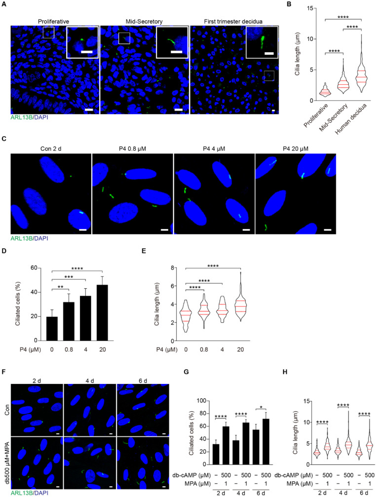
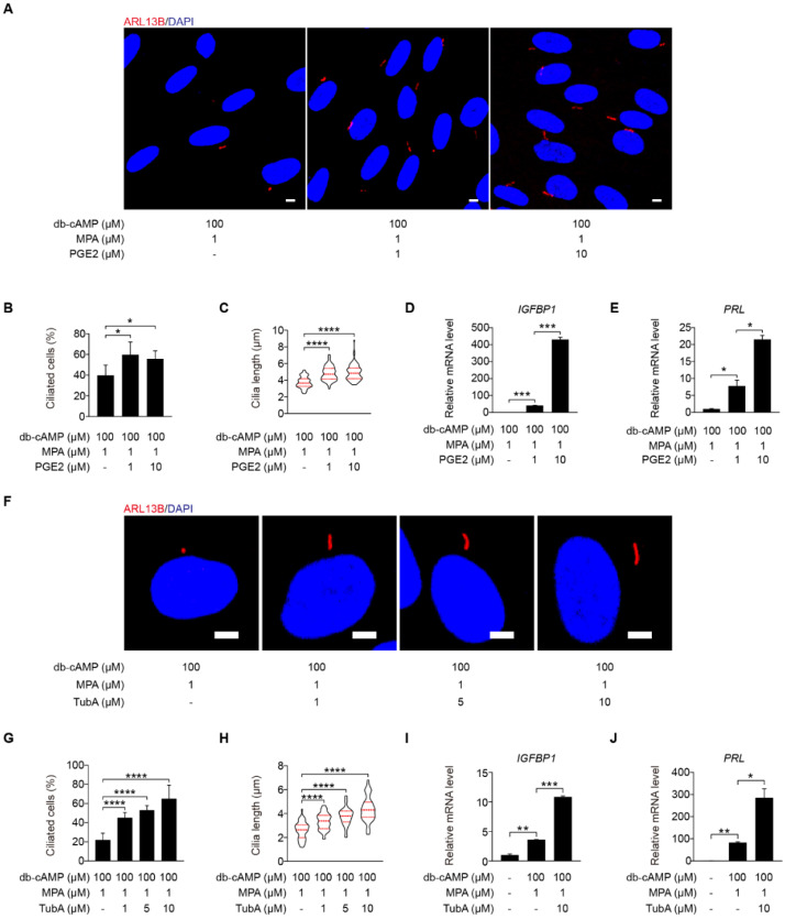
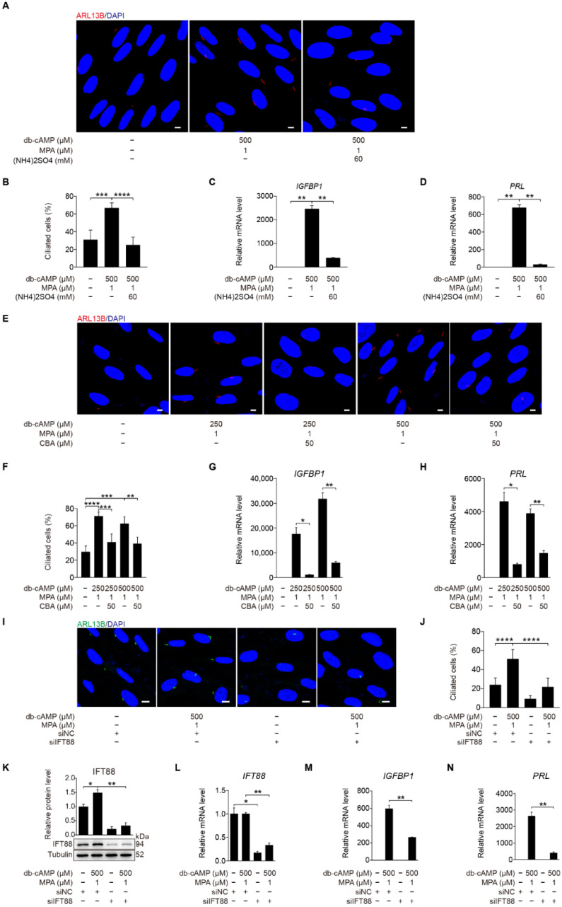
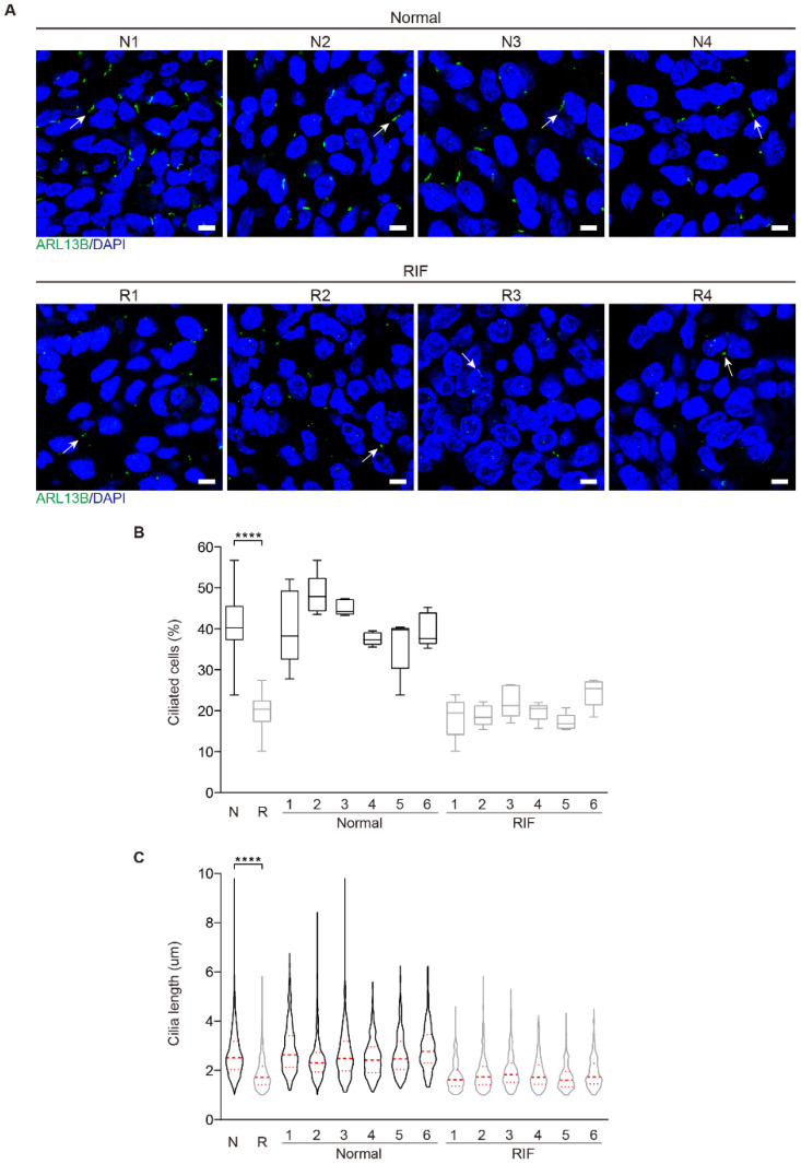
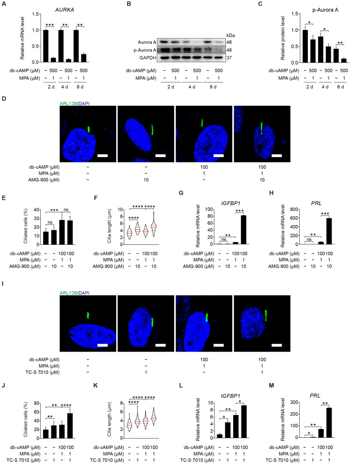
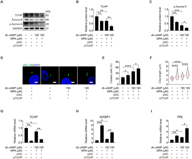
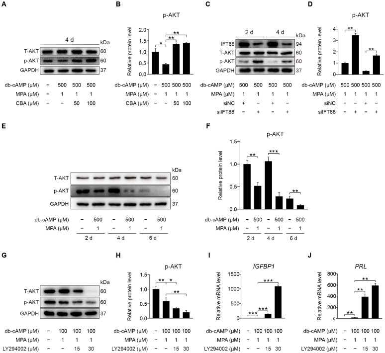
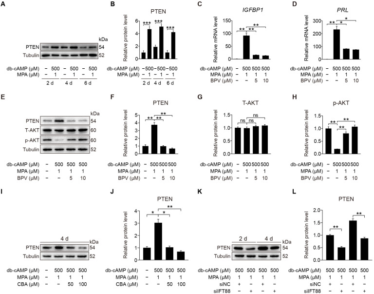
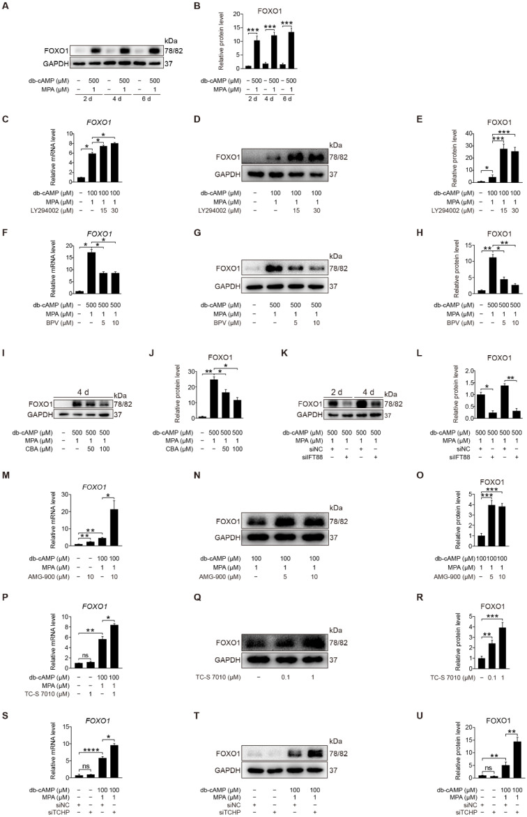
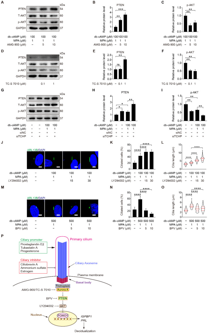
Endometrial decidualization plays a pivotal role during early pregnancy. Compromised decidualization has been tightly associated with recurrent implantation failure (RIF). Primary cilium is an antenna-like sensory organelle and acts as a signaling nexus to mediate Hh, Wnt, TGFβ, BMP, FGF, and Notch signaling. However, whether primary cilium is involved in human decidualization is still unknown. In this study, we found that primary cilia are present in human endometrial stromal cells. The ciliogenesis and cilia length are increased by progesterone during in vitro and in vivo decidualization. Primary cilia are abnormal in the endometrium of RIF patients. Based on data from both assembly and disassembly of primary cilia, it has been determined that primary cilium is essential to human decidualization. Trichoplein (TCHP)-Aurora A signaling mediates cilia disassembly during human in vitro decidualization. Mechanistically, primary cilium modulates human decidualization through PTEN-PI3K-AKT-FOXO1 signaling. Our study highlights primary cilium as a novel decidualization-related signaling pathway.

Keywords: AKT; Aurora A; decidualization; primary cilium.


Conflict of interest statement

The authors declare that they have no conflict of interest.

Figures

Figure 1
Figure 1
Primary cilia are present in both human endometrium in vivo and in decidual stromal cells in vitro. (A) ARL13B immunofluorescence in human endometrium during the proliferative, mid-secretory phase, and human first trimester decidua. ARL13B, a marker of the ciliary membrane, was used to visualize primary cilia (green). Nuclei were stained with DAPI (blue). Scale bars, 10 μm (main image) and 5 μm (magnified regions). (B) Cilia length in human endometrium and decidua. (C) ARL13B immunofluorescence in progesterone-treated stromal cells for 48 h. Scale bars, 5 μm. (D,E) Ciliated cells and cilia length were quantified in P4-treated stromal cells. (F) ARL13B immunofluorescence after stromal cells were induced for decidualization for 2, 4, and 6 days. Scale bars, 5 μm. (G,H) Ciliated cells and cilia length were quantified after stromal cells were induced for decidualization for 2, 4, and 6 days. In (A,C,F), the representative images of three biologically independent experiments are shown. In (B,E,H), these results are pooled from three independent experiments and presented as violin plots. The thick bar indicates the median and the dotted lines the first and third quartiles. In (B,D,E,G,H), data are presented as means ± SD from three independent experiments. * p < 0.05, ** p < 0.01, *** p < 0.001, and **** p < 0.0001 by two-tailed Student’s t test for comparing two groups or one-way ANOVA test for comparing more than two groups.
Figure 2
Figure 2
Primary cilia are critical for human decidualization. (A) ARL13B immunofluorescence in PGE2-treated stromal cells for 48 h. Scale bars, 5 μm. (B,C) Ciliated cells and cilia length were measured in PGE2-treated stromal cells for 48 h. (D,E) Effects of PGE2 treatment on relative mRNA levels of IGFBP1 and PRL under in vitro decidualization of stromal cells for 48 h. (F) ARL13B immunofluorescence in TubA-treated stromal cells for 48 h. Scale bars, 5 μm. (G,H) Ciliated cells and cilia length were measured in TubA-treated stromal cells for 48 h. (I,J) Relative mRNA levels of IGFBP1 and PRL in TubA-treated stromal cells for 48 h. In (A,F), the representative images of three biologically independent experiments are shown. In (C,H), the results are pooled from three independent experiments and presented as violin plots. The thick bar indicates the median and the dotted lines the first and third quartiles. In (BE,GJ), data are presented as means ± SD from three independent experiments. * p < 0.05, ** p < 0.01, *** p < 0.001, and **** p < 0.0001 by two-tailed Student’s t test for comparing two groups or one-way ANOVA test for comparing more than two groups.
Figure 3
Figure 3
Primary cilia are indispensable for human decidualization. (A) ARL13B immunofluorescence in (NH4)2SO4-treated stromal cells for 24 h under in vitro decidualization for 48 h. Scale bars, 5 μm. (B) Ciliated cells of stromal cells after (NH4)2SO4 treatment for 24 h under in vitro decidualization for 48 h. (C,D) Relative mRNA levels of IGFBP1 and PRL of stromal cells after (NH4)2SO4 treatment for 24 h under in vitro decidualization for 48 h. (E) Effects of CBA treatment for 24 h on ARL13B immunofluorescence under in vitro decidualization of stromal cells for 48 h. Scale bars, 5 μm. (F) Effects of CBA treatment for 24 h on the percentage of ciliated cells under in vitro decidualization of stromal cells for 48 h. (G,H) Effects of CBA treatment for 24 h on relative mRNA levels of IGFBP1 and PRL under in vitro decidualization of stromal cells for 48 h. (I) ARL13B immunofluorescence after stromal cells were transfected with siIFT88 for 24 h then in vitro decidualization for 48 h. Scale bars, 5 μm. (J,K) Ciliated cells and IFT88 protein levels after stromal cells were treated with siIFT88 for 24 h then in vitro decidualization for 48 h. (LN) Relative mRNA levels of IFT88, IGFBP1, and PRL after stromal cells were transfected with siIFT88 for 24 h followed by in vitro decidualization for 48 h. In (A,E,I), the representative images of three biologically independent experiments are shown. In (BD,FH,JN), data are presented as means ± SD from three independent experiments. * p < 0.05, ** p < 0.01, *** p < 0.001, and **** p < 0.0001 by two-tailed Student’s t test for comparing two groups or one-way ANOVA test for comparing more than two groups.
Figure 4
Figure 4
Aberrant primary cilia in the endometrium of RIF patients. (A) The representative images of ARL13B immunofluorescence in normal and RIF human endometrium. The representative ARL13B immunofluorescent images were shown from 4 control patients and 4 RIF patients. Primary cilia were identified with ARL13B antibody as green color. Nuclei were stained with DAPI (blue). Arrows, primary cilium; Scale bars, 5 μm. (B) Ciliated cells in normal and RIF human endometrium (N = 6, R = 6). The results are presented as median ± min/max whiskers in box plots. (C) Cilia length in normal and RIF human endometrium (N = 6, R = 6). The results are presented as violin plots, the thick bar indicates the median and the dotted lines the first and third quartiles. In (B,C), data are presented as means ± SD. **** p < 0.0001 by two-tailed Student’s t test for comparing two groups or one-way ANOVA test for comparing more than two groups.
Figure 5
Figure 5
Aurora A-mediated primary cilia disassembly inhibits human decidualization. (A) Relative mRNA level of AURKA during stromal cells decidualization for 2, 4, and 6 days. (B,C) Western blot and quantification of p-Aurora A (T288) during stromal cells decidualization for 2, 4, and 6 days. (D) ARL13B immunofluorescence in AMG-900-treated stromal cells for 48 h. Scale bars, 5 μm. (E,F) Ciliated cells and cilia length were measured in AMG-900-treated stromal cells for 48 h. (G,H) Relative mRNA levels of IGFBP1 and PRL in AMG-900-treated stromal cells for 48 h. (I) ARL13B immunofluorescence in TC-S 7010-treated stromal cells for 48 h. Scale bars, 5 μm. (J,K) Ciliated cells and cilia length were measured in TC-S 7010-treated stromal cells for 48 h. (L,M) Relative mRNA levels of IGFBP1 and PRL in TC-S 7010-treated stromal cells for 48 h. In (B,D,I), the representative images of three biologically independent experiments are shown. In (F,K), these results are pooled from three independent experiments and presented as violin plots. The thick bar indicates the median and the dotted lines the first and third quartiles. In (A,C,EH,JM), data are presented as means ± SD from three independent experiments. ns, (p > 0.05), * p < 0.05, ** p < 0.01, *** p < 0.001, and **** p < 0.0001 by two-tailed Student’s t test for comparing two groups or one-way ANOVA test for comparing more than two groups.
Figure 6
Figure 6
Trichoplein acts as the activator of Aurora A. (AC) Western blot and quantification of TCHP and p-Aurora A (T288) after stromal cells were transfected with siTCHP for 24 h then in vitro decidualization for 48 h. (D) ARL13B immunofluorescence after stromal cells were transfected with siTCHP for 24 h then in vitro decidualization for 48 h. Scale bars, 5 μm. (E,F) Ciliated cells and cilia length were analyzed after stromal cells were transfected with siTCHP for 24 h then in vitro decidualization for 48 h. (GI) Relative mRNA levels of TCHP, IGFBP1, and PRL after stromal cells were transfected with siTCHP for 24 h then in vitro decidualization for 48 h. In (A,D), the representative images of three biologically independent experiments are shown. In (F), the results are pooled from three independent experiments and presented as violin plots. The thick bar indicates the median and the dotted lines the first and third quartiles. In (B,C,EI), data are presented as means ± SD from three independent experiments. * p < 0.05, ** p < 0.01, *** p < 0.001, and **** p < 0.0001 by two-tailed Student’s t test for comparing two groups or one-way ANOVA test for comparing more than two groups.
Figure 7
Figure 7
Primary cilia negatively control AKT activation. (A,B) Western blot and quantification of p-AKT (S473) after stromal cells were treated with CBA for 24 h during in vitro decidualization for 4 days. (C,D) Western blot and quantification of p-AKT (S473) after stromal cells were treated with siIFT88 for 24 h then in vitro decidualization for 2 and 4 days. (E,F) Western blot and quantification of p-AKT (S473) in stromal cells during decidualization for 2, 4, and 6 days. (G,H) Western blot and quantification of p-AKT (S473) in LY294002-treated stromal cells for 48 h. (I,J) Relative mRNA levels of IGFBP1 and PRL in LY294002-treated stromal cells for 48 h. In (A,C,E,G), the representative images of three biologically independent experiments are shown. In (B,D,F,HJ), data are presented as means ± SD from three independent experiments. ns, (p > 0.05), * p < 0.05, ** p < 0.01, and *** p < 0.001 by two-tailed Student’s t test for comparing two groups or one-way ANOVA test for comparing more than two groups.
Figure 8
Figure 8
PTEN antagonizes AKT during stromal cells decidualization. (A,B) Western blot and quantification of PTEN in stromal cells during decidualization for 2, 4, and 6 days. (C,D) Relative mRNA levels of IGFBP1 and PRL in BPV-treated stromal cells for 48 h. (EH) Western blot and quantification of PTEN, T-AKT, and p-AKT (S473) in BPV-treated stromal cells for 48 h. (I,J) Western blot and quantification of PTEN after stromal cells were treated with CBA for 24 h during in vitro decidualization for 4 days. (K,L) Western blot and quantification of PTEN after stromal cells were treated with siIFT88 for 24 h then in vitro decidualization for 2 and 4 days. In (A,E,I,K), the representative images of three biologically independent experiments are shown. In (BD,FH,JL), data are presented as means ± SD from three independent experiments. ns, (p > 0.05), * p < 0.05, ** p < 0.01, and *** p < 0.001 by two-tailed Student’s t test for comparing two groups or one-way ANOVA test for comparing more than two groups.
Figure 9
Figure 9
Primary cilia modulate FOXO1 expression by suppressing AKT activity. (A,B) Western blot and quantification of FOXO1 in stromal cells during decidualization for 2, 4, and 6 days. (C) Relative mRNA level of FOXO1 in LY294002-treated stromal cells for 48 h. (D,E) Western blot and quantification of FOXO1 in LY294002-treated stromal cells for 48 h. (F) Relative mRNA level of FOXO1 in BPV-treated stromal cells for 48 h. (G,H) Western blot and quantification of FOXO1 in BPV-treated stromal cells for 48 h. (I,J) Western blot and quantification of FOXO1 after stromal cells were treated with CBA for 24 h during in vitro decidualization for 4 days. (K,L) Western blot and quantification of FOXO1 after stromal cells were treated with siIFT88 for 24 h then in vitro decidualization for 2 and 4 days. (M) Relative mRNA level of FOXO1 in AMG-900-treated stromal cells for 48 h. (N,O) Western blot and quantification of FOXO1 in AMG-900-treated stromal cells for 48 h. (P) Relative mRNA level of FOXO1 in TC-S 7010-treated stromal cells for 48 h. (Q,R) Western blot and quantification of FOXO1 in TC-S 7010-treated stromal cells for 48 h. (S) Relative mRNA level of FOXO1 after stromal cells were treated with siTCHP for 24 h then in vitro decidualization for 2 days. (T,U) Western blot and quantification of FOXO1 after stromal cells were treated with siTCHP for 24 h then in vitro decidualization for 2 days. In (A,D,G,I,K,N,Q,T), the representative images of three biologically independent experiments are shown. In (B,C,E,F,H,M,J,L,M,O,P,R,S,U), data are presented as means ± SD from three independent experiments. ns, (p > 0.05), * p < 0.05, ** p < 0.01, *** p < 0.001 and **** p < 0.0001 by two-tailed Student’s t test for comparing two groups or one-way ANOVA test for comparing more than two groups.
Figure 10
Figure 10
Trichoplein-Aurora A-mediated cilia disassembly contributes to PTEN-AKT-FOXO1 signaling. (AC) Western blot and quantification of PTEN and p-AKT in AMG-900-treated stromal cells for 48 h. (DF) Western blot and quantification of PTEN and p-AKT in TC-S 7010-treated stromal cells for 48 h. (GI) Western blot and quantification of PTEN and p-AKT after stromal cells were transfected with siTCHP for 24 h then in vitro decidualization for 48 h. (J) ARL13B immunofluorescence in LY294002-treated stromal cells for 48 h. Scale bars, 5 μm. (K,L) Ciliated cells and cilia length were measured in LY294002-treated stromal cells for 48 h. (M) ARL13B immunofluorescence in BPV-treated stromal cells for 48 h. Scale bars, 5 μm. (N,O) Ciliated cells and cilia length were measured in BPV-treated stromal cells for 48 h. (P) Model of primary cilia function in stromal cells decidualization. In (A,D,G,J,M), the representative images of three biologically independent experiments are shown. In (L,O), the results are pooled from three independent experiments and presented as violin plots. The thick bar indicates the median and the dotted lines in the first and third quartiles. In (B,C,E,F,H,I,K,L,N,O), data are presented as means ± SD from three independent experiments. ns, (p > 0.05), * p < 0.05, ** p < 0.01, *** p < 0.001, and **** p < 0.0001 by two-tailed Student’s t test for comparing two groups or one-way ANOVA test for comparing more than two groups.

References

    1. Ramathal C.Y., Bagchi I.C., Taylor R.N., Bagchi M.K. Endometrial decidualization: Of mice and men. Semin. Reprod. Med. 2010;28:17–26. doi: 10.1055/s-0029-1242989. - DOI - PMC - PubMed
    1. Zhang X.H., Liang X., Liang X.H., Wang T.S., Qi Q.R., Deng W.B., Sha A.G., Yang Z.M. The mesenchymal-epithelial transition during in vitro decidualization. Reprod. Sci. 2013;20:354–360. doi: 10.1177/1933719112472738. - DOI - PMC - PubMed
    1. Gellersen B., Brosens J. Cyclic AMP and progesterone receptor cross-talk in human endometrium: A decidualizing affair. J. Endocrinol. 2003;178:357–372. doi: 10.1677/joe.0.1780357. - DOI - PubMed
    1. Matsumoto H., Zhao X., Das S.K., Hogan B.L., Dey S.K. Indian hedgehog as a progesterone-responsive factor mediating epithelial-mesenchymal interactions in the mouse uterus. Dev. Biol. 2002;245:280–290. doi: 10.1006/dbio.2002.0645. - DOI - PubMed
    1. Sonderegger S., Pollheimer J., Knofler M. Wnt signalling in implantation, decidualisation and placental differentiation--review. Placenta. 2010;31:839–847. doi: 10.1016/j.placenta.2010.07.011. - DOI - PMC - PubMed

Substances