. 2022 Dec 9;23(24):15591.
doi: 10.3390/ijms232415591.

Myxococcus xanthus Encapsulin as a Promising Platform for Intracellular Protein Delivery

Affiliations

Myxococcus xanthus Encapsulin as a Promising Platform for Intracellular Protein Delivery

Anna N Gabashvili et al. Int J Mol Sci. .

Abstract

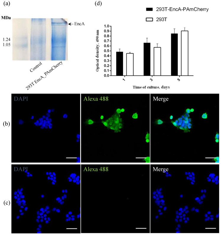
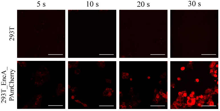
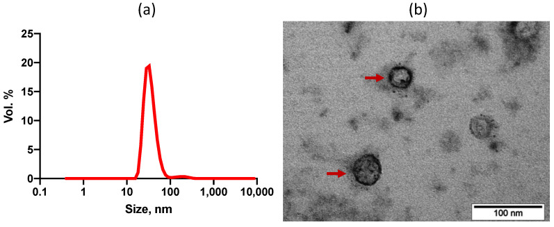
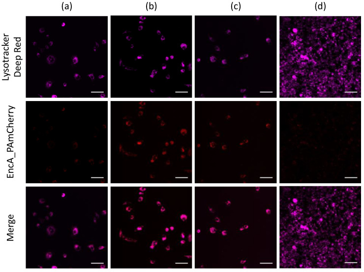
Introducing a new genetically encoded material containing a photoactivatable label as a model cargo protein, based on Myxococcus xanthus (Mx) encapsulin system stably expressed in human 293T cells. Encapsulin from Mx is known to be a protein-based container for a ferritin-like cargo in its shell which could be replaced with an exogenous cargo protein, resulting in a modified encapsulin system. We replaced Mx natural cargo with a foreign photoactivatable mCherry (PAmCherry) fluorescent protein and isolated encapsulins, containing PAmCherry, from 293T cells. Isolated Mx encapsulin shells containing photoactivatable label can be internalized by macrophages, wherein the PAmCherry fluorescent signal remains clearly visible. We believe that a genetically encoded nanocarrier system obtained in this study, can be used as a platform for controllable delivery of protein/peptide therapeutics in vitro.

Keywords: encapsulins; fluorescence; photoactivatable label; protein delivery.


Conflict of interest statement

The authors declare no conflict of interest.

Figures

Figure 1
Figure 1
(a) Coomasie-stained BN-PAGE gels loaded with 293T and 293T EncA_PAmCherry cell lysates. Arrow and dotted line indicate the band corresponding to Mx EncA protein shells; (b) 293T EncA_PAmCherry and (c) 293T cells stained with Alexa Fluor 488 anti-DYKDDDDK Tag antibody (green fluorescence signal). Nuclei were counterstained with DAPI (blue fluorescence signal). Laser scanning confocal microscopy, Nikon Eclipse Ti2, scale bars are 50 μm; (d) Influence of the presence of a genetically encoded label on viability and proliferation of 293T EncA_PAmCherry cells. Optical density is proportional to the number of living cells. The numbers of living cells were not significantly different in 293T EncA_PAmCherry and 293T cells. The data are shown as the mean + S.D. of three independent experiments, p values were calculated using a one-tailed t-test, assuming unequal variances.
Figure 2
Figure 2
293T (upper panel) and 293T EncA_PAmCherry (lower panel) cells were imaged after irradiation with 405 nm laser for 5 s, 10 s, 20 s and 30 s followed by 561 nm excitation. Laser scanning confocal microscopy, Nikon Eclipse Ti2, scale bars are 50 μm.
Figure 3
Figure 3
(a) Dynamic light scattering analysis of EncA_PAmCherry (34 ± 4 nm; PDI 0,22). Hydrodynamic size of Mx encapsulins, volume distribution. (b) Bright-field TEM image of Uranyless -stained EncA_PAmCherry, red arrows indicate Mx encapsulins protein shells, scale bar 100 nm.
Figure 4
Figure 4
Confocal imaging of RAW 264.7 cells after (a) 15 min, (b) 1 h, (c) 2 h and (d) 24 h of incubation with isolated EncA_PAmCherry. RAW 264.7 cells were imaged after irradiation with 405 nm laser for 30 s followed by 561 nm excitation of PAmCherry label (red fluorescence signal). Lysosomes were stained with LysoTracker Deep Red dye (purple fluorescence signal). Laser scanning confocal microscopy, Nikon Eclipse Ti2 and scale bars are 50 μm.

References

    1. Yu M., Wu J., Shi J., Farokhzad O.C. Nanotechnology for Protein Delivery: Overview and Perspectives. J. Control. Release. 2016;240:24–37. doi: 10.1016/j.jconrel.2015.10.012. - DOI - PMC - PubMed
    1. Dean S.N., Turner K.B., Medintz I.L., Walper S.A. Targeting and Delivery of Therapeutic Enzymes. Ther. Deliv. 2017;8:577–595. doi: 10.4155/tde-2017-0020. - DOI - PubMed
    1. Akash M.S.H., Rehman K., Chen S. Pluronic F127-Based Thermosensitive Gels for Delivery of Therapeutic Proteins and Peptides. Polym. Rev. 2014;54:573–597. doi: 10.1080/15583724.2014.927885. - DOI
    1. Fletcher N.A., Babcock L.R., Murray E.A., Krebs M.D. Controlled Delivery of Antibodies from Injectable Hydrogels. Mater. Sci. Eng. C. 2016;59:801–806. doi: 10.1016/j.msec.2015.10.096. - DOI - PubMed
    1. Ribeiro M.P., Morgado P.I., Miguel S.P., Coutinho P., Correia I.J. Dextran-Based Hydrogel Containing Chitosan Microparticles Loaded with Growth Factors to Be Used in Wound Healing. Mater. Sci. Eng. C. 2013;33:2958–2966. doi: 10.1016/j.msec.2013.03.025. - DOI - PubMed

MeSH terms

Substances

LinkOut - more resources