. 2022 Dec 12;23(24):15742.
doi: 10.3390/ijms232415742.

The J Domain of Sacsin Disrupts Intermediate Filament Assembly

Affiliations

The J Domain of Sacsin Disrupts Intermediate Filament Assembly

Afrooz Dabbaghizadeh et al. Int J Mol Sci. .

Abstract

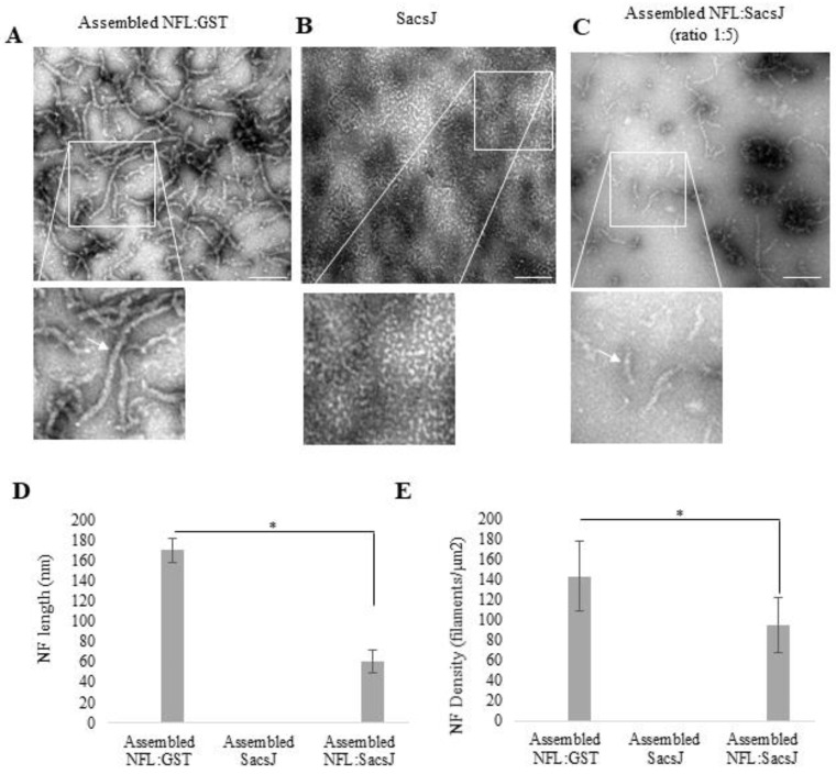
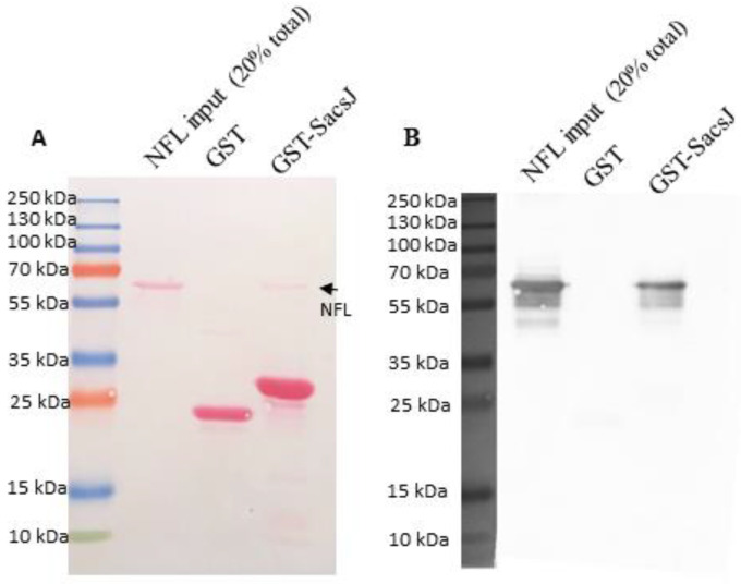
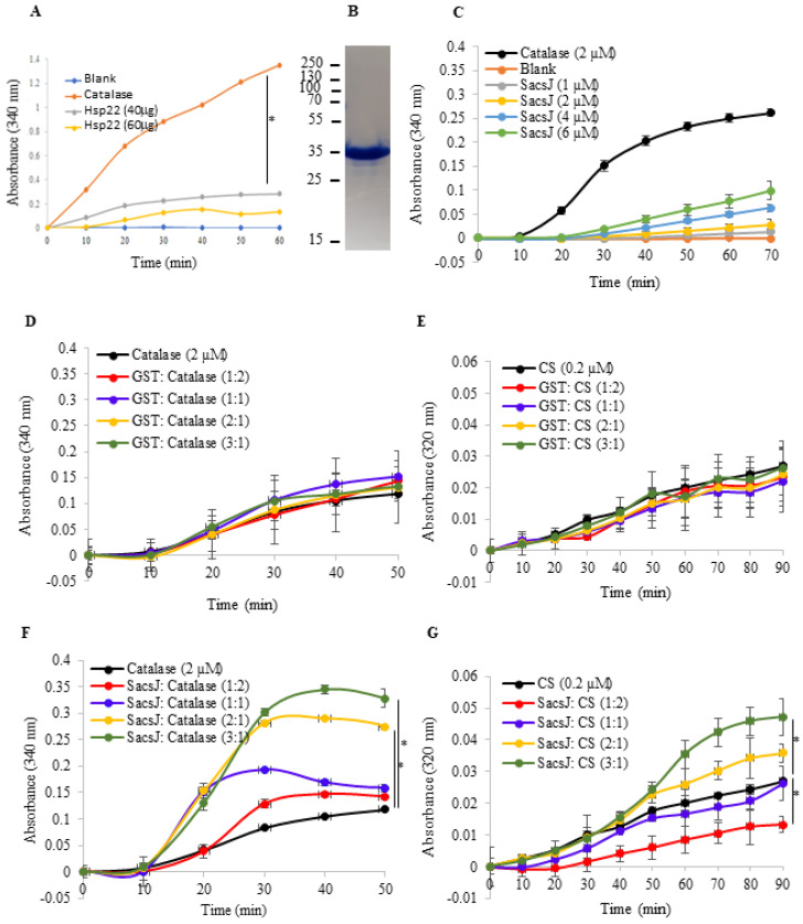
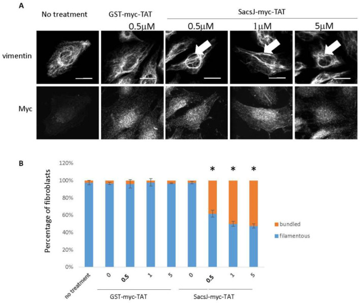
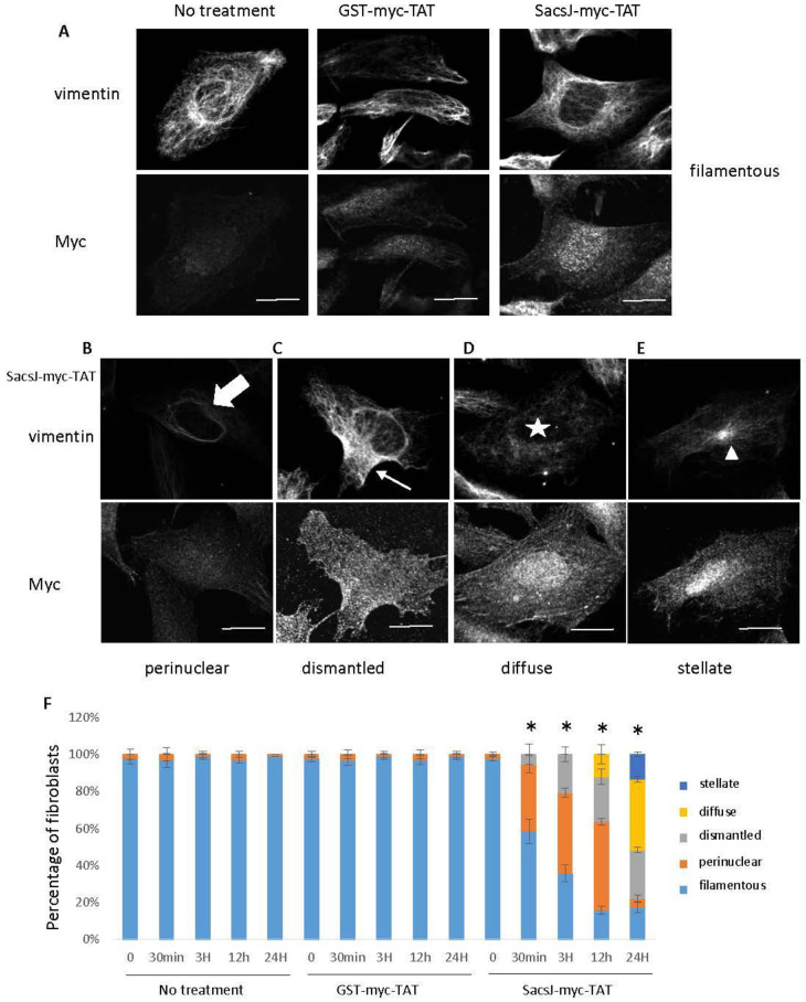
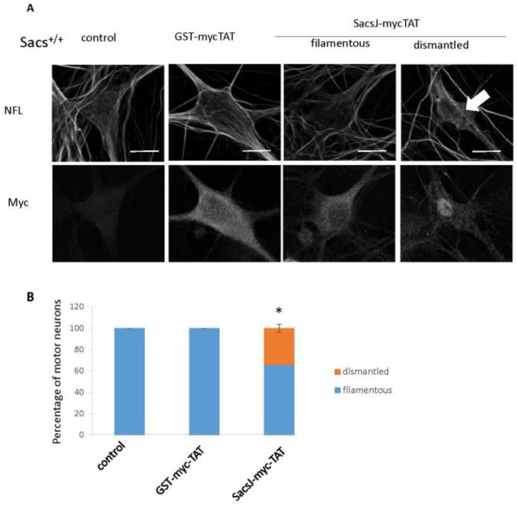
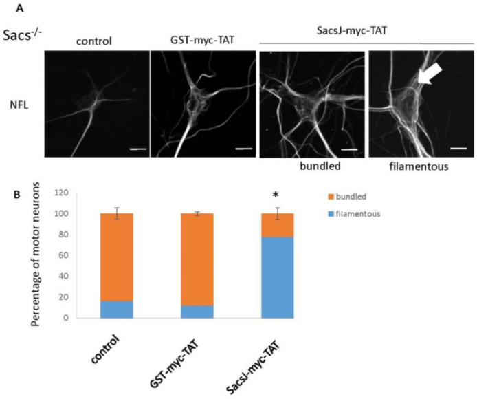
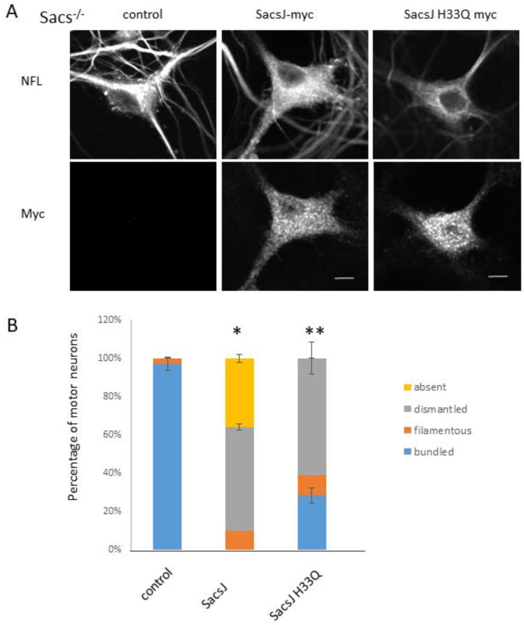
Autosomal Recessive Spastic Ataxia of the Charlevoix Saguenay (ARSACS) is caused by mutation in the SACS gene resulting in loss of function of the protein sacsin. A key feature is the formation of abnormal bundles of neurofilaments (NF) in neurons and vimentin intermediate filaments (IF) in cultured fibroblasts, suggesting a role of sacsin in IF homeostasis. Sacsin contains a J domain (SacsJ) homologous to Hsp40, that can interact with Hsp70 chaperones. The SacsJ domain resolved NF bundles in cultured Sacs-/- neurons. Having studied the mechanism using NF assembled in vitro from purified NF proteins, we report that the SacsJ domain interacts with NF proteins to disassemble NFL filaments, and to inhibit their initial assembly. A cell-penetrating peptide derived from this domain, SacsJ-myc-TAT was efficient in disassembling NF bundles in cultured Sacs-/- motor neurons, restoring the NF network; however, there was some loss of vimentin IF and NF in cultured Sacs+/+ fibroblasts and motor neurons, respectively. These results suggest that sacsin through its SacsJ domain is a key regulator of NF and vimentin IF networks in cells.

Keywords: J domain; ataxia; chaperone; intermediate filaments; motor neuron; neurofilament; vimentin.


Conflict of interest statement

The authors declare no conflict of interest. The authors declare no competing financial interest.

Figures

Figure 1
Figure 1
SacsJ disassembled NFL filaments in vitro. Shown are representative TEM images of filaments assembled from purified NFL (A). Assembled NFL filaments were incubated with or without SacsJ for 1 h at 37 °C and observed by TEM. Representative TEM images of filamentous NFL (A), SacsJ alone (B) or filamentous NFL co-incubated with SacsJ at a molar ratio of 1:5 (C). Inserts show enlargements of TEM images focused on NFL filaments (arrow). Scale Bar: 40 nm. Quantitation of NFL average length (D) and filament density (E) with or without incubation with SacsJ shows the significant decrease in NFL length and density (filaments/µm2). * p < 0.05 vs. NFL alone; one-way ANOVA, HSD Tuckey post hoc analysis (n = 3).
Figure 2
Figure 2
SacsJ impaired NFL assembly in vitro. (AE) TEM of negatively stained preparations of assembled NFL dialyzed alone (A) or in the presence of different molar ratios of NFL to GST-SacsJ: 1:1 (B,C), 1:5 (D), and 1:10 (E). Scale bars: 100 nm. SacsJ prevented formation of NFL filaments (arrow). (F) Quantitation of NFL filament density: Incubation with GST-SacsJ significantly reduced filament density in a concentration-dependent fashion (filaments/µm2). * p < 0.05 vs. NFL alone, one-way ANOVA (n = 3). (G) SDS-PAGE of an in vitro sedimentation assay of NFL with or without SacsJ after assembly at 4 °C overnight at NFL:SacsJ ratio of 1:6. The supernatant (S) and pellet (P) fractions were analyzed by SDS-PAGE followed by Coomassie Blue staining. NFL and SacsJ bands are indicated by arrows and molecular weight is on the left.
Figure 3
Figure 3
SacsJ directly interacts with NFL in vitro. (A) GST pulldown assay of soluble recombinant NFL with GST-SacsJ, or GST as control. (A) Ponceau red staining of the nitrocellulose membrane transferred from SDS-PAGE. Lanes are: NFL input—20% of total amount of NFL used in the pulldown assay; GST: GST incubated with NFL; GST-SacsJ: GST-SacsJ incubated with NFL. Bands stained by Ponceau red in each lane are NFL, GST, GST–SacsJ and NFL (small arrow). (B) Immunolabelling of the nitrocellulose membrane shown in (A) using anti-NFL antibody (NR4, 1/1000) demonstrating that the small band identified in (A) using GST-SacsJ as bait is indeed NFL.
Figure 4
Figure 4
SacsJ domain did not act as a chaperone to reduce heat-denaturation of catalase or citrate synthase in an in vitro assay. (A) Absorbance at 340 nm of catalase (2 µM) incubated with or without Hsp22 used as a positive control (40 or 60 µg) heated at 60 °C. (B) Coomassie brilliant blue staining of a SDS-PAGE of purified recombinant SacsJ protein used in these assays. (C) Absorbance at 340 nm, as a function of time in an assay for heat-denaturation of catalase (2 µM) at 60 °C compared to heat-induced denaturation in the presence of increasing concentrations of SacsJ-GST (1–6 µM). (D,E) Absorbance at 340 nm of catalase (2 µM) or at 320 nm citrate synthase (CS) (0.2 µM) incubated with or without GST (1–6 µM) heated at 60 °C for catalase or 45 °C for CS. (F,G) Absorbance at 340 nm and 320 nm of catalase (2 µM) or citrate synthase (CS) (0.2 µM) incubated with or without SacsJ-GST (1–6 µM) heated at 60 °C for catalase or 45 °C for CS. * p < 0.05 vs. catalase or CS alone, one-way ANOVA, HSD Tuckey post hoc analysis (n = 3).
Figure 5
Figure 5
The cell-permeant peptide, Sacs-myc-TAT, induced step-wise disruption of the vimentin IF network in MCH74 fibroblasts. (A) Representative 3-dimensional reconstructions of Z-stack confocal images of MCH74 fibroblasts double labelled with anti-myc (rabbit anti-myc) and anti-vimentin (V9 mouse monoclonal) treated with increasing concentrations of Sacs-myc-TAT as indicated (0.5 to 5 µM) or control peptide GST-myc-TAT. Treatment with Sacs-myc-TAT for 30 min resulted in nuclear rings of bundled vimentin IF (large arrow). Scale bar: 20 µm. (B) Quantitation of the percentage of fibroblasts presenting circum-nuclear IF bundles or finely distributed IF when treated with increasing concentrations of Sacs-myc-TAT (0.5 to 5 µM) showing an increase in the percentage of cells with IF concentrated surrounding the nucleus. * p < 0.05 vs. NFL alone, one-way ANOVA (n = 3). Note the diffuse distribution of labelling of the myc tag on SacsJ.
Figure 6
Figure 6
Over time, SacsJ-myc-TAT treatment resulted in the disassembly of vimentin IF in normal Sacs+/+ (MCH74) fibroblasts. (A) Representative 3-dimensional reconstructions of Z-stack confocal images of fibroblasts double labelled with anti-myc (rabbit anti-myc) and anti-vimentin (V9 mouse monoclonal). Scale bar: 20 µm. (BE) MCH74 fibroblasts were treated with GST-myc-TAT control or SacsJ-myc-TAT (0.5 µM) for 30 min, 3 h, 12 h or 24 h and showed time-dependent phenotypes: perinuclear rings of vimentin ((B), large arrow), dismantled vimentin network ((C), small arrow), diffuse vimentin labelling ((D), star), and appearance at a stellate accumulation ((E), arrowhead). (F) Quantitation of the percentage of fibroblasts presenting those phenotypes over duration of SacsJ-myc-TAT treatment. * p < 0.05 vs. time-matched no treatment or treated with GST-myc-TAT (0.5 µM) one-way ANOVA, HSD Tuckey post hoc analysis (n = 3).
Figure 7
Figure 7
The cell-permeant peptide, SacsJ-myc-TAT, induced the disassembly of NF in Sacs+/+ motor neuron in culture. (A) Representative 3-dimensional reconstructions of Z-stack confocal images of double labelling with anti-myc (rabbit anti-myc) and anti-NFL in Sacs+/+ 6 week-old murine spinal cord-DRG cultures showing the NF network and distribution of myc-TAT peptides in motor neurons. Cultures were treated with SacsJ-myc-TAT (0.5 µM) or GST-myc-TAT control peptide for 30 min and compared to untreated cultures. SacsJ-myc-TAT dismantled the endogenous NF network (large arrow). Scale bar: 20 μm. (B) Quantitation of the percentage of motor neurons presenting a filamentous or dismantled NF network when treated with Sacs-myc-TAT (0.5 μM). * p < 0.05 vs. no treatment or treated with GST-myc-TAT (0.5 µM) using a one-way ANOVA, HSD Tuckey post hoc analysis (n = 3).
Figure 8
Figure 8
The cell-penetrating peptide, SacsJ-myc-TAT, resolved NFL bundles in Sacs−/− motor neuron in culture. (A) Representative 3-dimensional reconstructions of Z-stack confocal images of motor neurons in Sacs−/− 6 week-old spinal cord-DRG cultures double labelled with anti-myc (rabbit anti-myc) and anti-NFL to show the NF network and SacsJ-myc-TAT distribution in motor neurons. Cultures were treated with SacsJ-myc-TAT (0.5 µM) or GST-myc-TAT control peptide for 30 min and compared to untreated cultures. Treatment with SacsJ-myc-TAT resolved the NF bundles (large arrow). Scale bar: 20 µm. (B) Quantitation of the percentage of motor neurons presenting a filamentous or bundled NF network when treated with Sacs-myc-TAT (0.5 µM). * p < 0.05 vs. no treatment or treated with GST-myc-TAT (0.5 µM) using a one-way ANOVA, HSD Tuckey post hoc analysis (n = 3).
Figure 9
Figure 9
SacsJ acts independently to disassemble NF, but requires Hsp70 to degrade NFL. SacsJ-myc or SacsJH33Q-myc, a variant unable to bind HSP70, were expressed in motor neurons in Sacs−/− 6 week-old spinal cord-DRG cultures. (A) Representative 3-dimensional reconstructions of Z-stack confocal images of motor neurons double labelled with anti-myc (rabbit anti-myc) and anti-NFL to show the NF network and expression of NFL. The NF network was dismantled or absent when SacsJ-myc was expressed while SacsJH33Q-myc only dismantled NF network. Scale bar: 10 µm. (B) Quantitation of the percentage of motor neurons presenting a filamentous, bundled, dismantled or no NF network when expressing SacsJ-myc or SacsJH33Q-myc. * p < 0.05 vs. control, ** p < 0.05 vs. SacsJ using a one-way ANOVA, HSD Tuckey post hoc analysis (n = 3).

References

    1. Vingolo E.M., Di Fabio R., Salvatore S., Grieco G., Bertini E., Leuzzi V., Nesti C., Filla A., Tessa A., Pierelli F., et al. Myelinated retinal fibers in autosomal recessive spastic ataxia of Charlevoix-Saguenay. Eur. J. Neurol. 2011;18:1187–1190. doi: 10.1111/j.1468-1331.2010.03335.x. - DOI - PubMed
    1. Garcia-Martin E., Pablo L.E., Gazulla J., Polo V., Ferreras A., Larrosa J.M. Retinal nerve fibre layer thickness in ARSACS: Myelination or hypertrophy? Br. J. Ophthalmol. 2013;97:238–241. doi: 10.1136/bjophthalmol-2012-302309. - DOI - PMC - PubMed
    1. Girard M., Larivière R., Parfitt D.A., Deane E.C., Gaudet R., Nossova N., Blondeau F., Prenosil G., Vermeulen E.G.M., Duchen M.R., et al. Mitochondrial dysfunction and Purkinje cell loss in autosomal recessive spastic ataxia of Charlevoix-Saguenay (ARSACS) Proc. Natl. Acad. Sci. USA. 2012;109:1661–1666. doi: 10.1073/pnas.1113166109. - DOI - PMC - PubMed
    1. Baets J., Deconinck T., Smets K., Goossens D., Van den Bergh P., Dahan K., Schmedding E., Santens P., Rasic V.M., Van Damme P., et al. Mutations in SACS cause atypical and late-onset forms of ARSACS. Neurology. 2010;75:1181–1188. doi: 10.1212/WNL.0b013e3181f4d86c. - DOI - PubMed
    1. Bouchard J.P., Barbeau A., Bouchard R., Bouchard R.W. Electromyography and nerve conduction studies in Friedreich’s ataxia and autosomal recessive spastic ataxia of Charlevoix-Saguenay (ARSACS) Can. J. Neurol. Sci. 1979;6:185–189. doi: 10.1017/S0317167100119614. - DOI - PubMed

Supplementary concepts

LinkOut - more resources