. 2022 Dec 13;23(24):15784.
doi: 10.3390/ijms232415784.

Neural Stem Cells Overexpressing Arginine Decarboxylase Improve Functional Recovery from Spinal Cord Injury in a Mouse Model

Affiliations

Neural Stem Cells Overexpressing Arginine Decarboxylase Improve Functional Recovery from Spinal Cord Injury in a Mouse Model

Yu Mi Park et al. Int J Mol Sci. .

Abstract

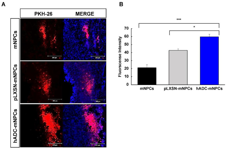
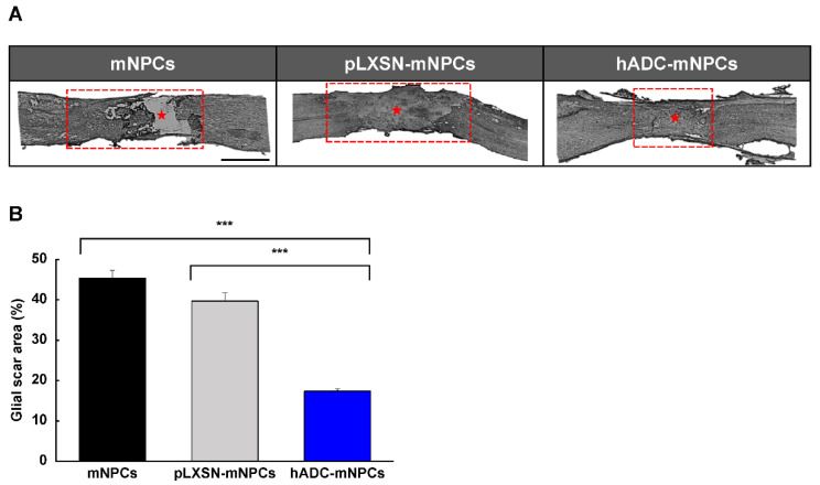
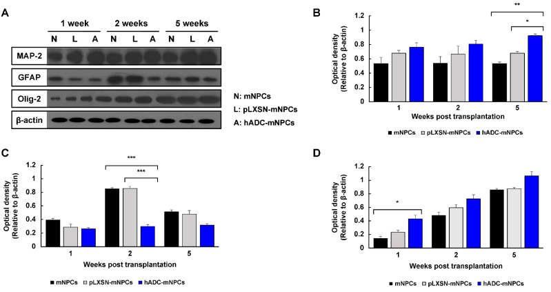
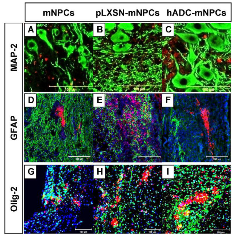
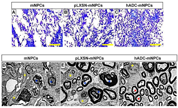
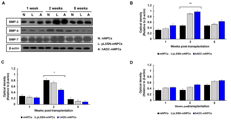
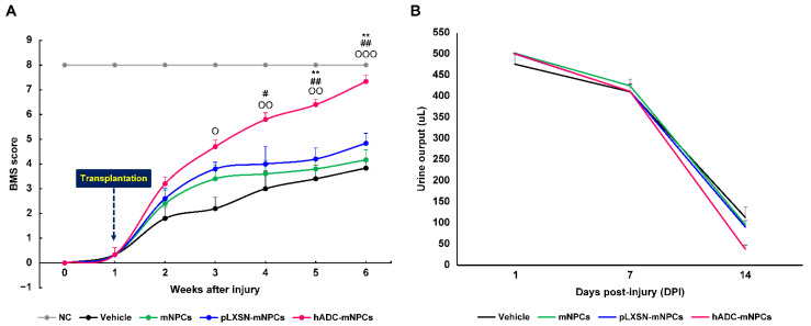
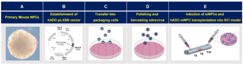
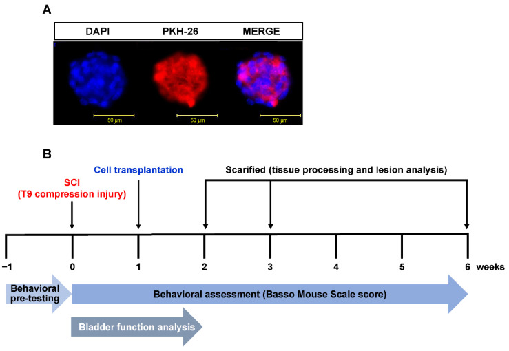
Current therapeutic strategies for spinal cord injury (SCI) cannot fully facilitate neural regeneration or improve function. Arginine decarboxylase (ADC) synthesizes agmatine, an endogenous primary amine with neuroprotective effects. Transfection of human ADC (hADC) gene exerts protective effects after injury in murine brain-derived neural precursor cells (mNPCs). Following from these findings, we investigated the effects of hADC-mNPC transplantation in SCI model mice. Mice with experimentally damaged spinal cords were divided into three groups, separately transplanted with fluorescently labeled (1) control mNPCs, (2) retroviral vector (pLXSN)-infected mNPCs (pLXSN-mNPCs), and (3) hADC-mNPCs. Behavioral comparisons between groups were conducted weekly up to 6 weeks after SCI, and urine volume was measured up to 2 weeks after SCI. A subset of animals was euthanized each week after cell transplantation for molecular and histological analyses. The transplantation groups experienced significantly improved behavioral function, with the best recovery occurring in hADC-mNPC mice. Transplanting hADC-mNPCs improved neurological outcomes, induced oligodendrocyte differentiation and remyelination, increased neural lineage differentiation, and decreased glial scar formation. Moreover, locomotor and bladder function were both rehabilitated. These beneficial effects are likely related to differential BMP-2/4/7 expression in neuronal cells, providing an empirical basis for gene therapy as a curative SCI treatment option.

Keywords: agmatine; arginine decarboxylase; axonal re-myelination; cell transplantation; functional recovery; gene therapy; glial scar; neural progenitor cells; neurogenesis; spinal cord injury.


Conflict of interest statement

The authors declare no conflict of interest.

Figures

Figure 1
Figure 1
Migration and survival of mNPCs around the lesion site at 1 week after cell transplantation following SCI. (A) A higher number of PKH-26-labeled hADC-mNPCs grafted found around the lesion area (T9) than there were mNPCs and pLXSN-mNPCs at 1 week after cell transplantation. (B) More PKH-26-labeled hADC-mNPCs (60% fluorescence intensity) were present around the lesion area (T9) than there were mNPCs (20%) and pLXSN-mNPCs (40%). Data are presented as means ± standard error of mean (SEM). (n = 5 per sample, * p < 0.05, *** p < 0.001).
Figure 2
Figure 2
hADC overexpression in mNPCs reduces glial scar volume 2 weeks after transplantation into compression-lesioned spinal cords of adult mice. (A) Immunohistochemistry of longitudinal spinal cord sections stained with antibodies against GFAP at T8, T9, and T10 at 2 weeks after transplantation of hADC overexpressing mNPCs (hADC-mNPCs), empty retroviral overexpression mNPCs (pLXSN-mNPCs), or mNPCs alone. The red box was the site of the glial scar lesion, and the middle of the glial scar is indicated by a red asterisk. Scale bar = 5 mm. (B) Lesion areas are significantly larger in mice with mNPC transplantation (45% of the area shown in part A) than transplantations of pLXSN-mNPCs (40% of the area shown in A) and hADC-mNPCs (19% of the area shown in A). Data are presented as means ± SEM. (n = 5 per sample, *** p < 0.001).
Figure 3
Figure 3
Western blotting of MAP-2, GFAP, Olig-2, and β-actin from T8, T9 (lesion area), and T10 at 1, 2, and 5 weeks after cell transplantation. (A) Representative Western blots of MAP-2, GFAP, Olg-2, and β-actin. (B) Quantification of MAP-2, showing that hADC-mNPC mice had significantly higher expression than pLXSN-mNPC or mNPC mice at 5 weeks post-transplantation. (C) Quantification of GFAP, showing a significant decrease in the hADC-mNPC group compared with the pLXSN-mNPC and mNPC groups at 2 weeks post-transplantation. (D) Quantification of Olig-2, showing a significant increase in the hADC-mNPC group compared with the pLXSN-mNPC and mNPC groups at 1 week post-transplantation. Data are presented as the mean ± SEM. (n = 5 per sample, * p < 0.05, ** p < 0.01, *** p < 0.001).
Figure 4
Figure 4
Double immunohistochemistry of PKH-26-labeled mNPCs, pLXSN-mNPCs, and hADC-mNPCs, along with the marker microtubule associated protein-2 (MAP-2), fibrillary acidic protein (GFAP), and Olig-2 around lesion sites after SCI. (AC) Immunofluorescence staining of MaP-2 (green) in PKH-26-labeled mNPCs, pLXSN-mNPCs and hADC-mNPCs (red) at 5 weeks post-transplantation. (DF) Immunofluorescence staining of GFAP (green) in PKH-26-labeled mNPCs, pLXSN-mNPCs and hADC-mNPCs (red) at 1 week post-transplantation. DAPI staining (blue) indicating the nuclei. (GI) Immunofluorescence staining of Olig-2 (green) in PKH-26-labeled mNPCs, pLXSN-mNPCs, and hADC-mNPCs (red) at 1 week post-transplantation. DAPI staining (blue) indicating the nuclei. Scale bar = 100 μm (MAP-2 and Olig-2), 200 μm (GFAP).
Figure 5
Figure 5
Luxol fast blue staining of T9 spinal cord sections obtained from the mNPC (A), pLXSN-mNPC (B), and hADC-mNPC (C) transplantation groups at 6 weeks after SCI. The hADC-mNPC group (C) had smaller cystic cavities and more myelin sheaths than the other two groups (A,B). Scale bar = 20 μm. Transmission electron microscopy (TEM, 10,000×) of the lumbar section where mNPCs (D), pLXSN-mNPCs (E), and hADC-mNPCs (F) were transplanted after SCI. Transplanted hADC-mNPCs promoted axon remyelination at the lesion site. Red stars indicate remyelinated or mature axons in the hADC-mNPC group, while blue and yellow stars indicate degenerating myelinated axons and microglia/macrophage cells, respectively.
Figure 6
Figure 6
Western blot analysis of BMP-2, BMP-4, BMP-7, and β-actin from T8, T9, and T10 at 1, 2, and 5 weeks after cell transplantation. (A) Representative Western blots of BMP-2, BMP-4, BMP-7, and β-actin. (B) Quantification of BMP-2 showing a significant increase in hADC-mNPC mice compared with pLXSN-mNPC and mNPC mice at 2 weeks post-transplantation. (C) Quantification of BMP-4 showing a significant decrease in hADC-mNPC mice compared with pLXSN-mNPC and mNPC mice at 2 weeks post-transplantation. (D) Quantification of BMP-7 demonstrating a lack of significant difference across the three groups, despite slightly higher expression in the hADC-mNPC group at 1, 2 and 5 weeks post-transplantation. Data are presented as means ± SEM. (n = 5 per sample, * p < 0.05, ** p < 0.01).
Figure 7
Figure 7
Mice with hADC-overexpressing mNPCs (hADC-mNPCs) exhibit improved motor performances and bladder function recovery. (A) Basso Mouse Scale (BMS) score up to 6 weeks after SCI, demonstrating continuous improvement in the hADC-mNPC group compared with other groups at 4, 5, and 6 weeks. (B) Comparison of bladder residual urine volumes up to 14 days post-SCI, showing that levels were lower in hADC-mNPC mice than in other mice. Data are presented as means ± SEM. (n = 10−40 per group, O p < 0.05, OO p < 0.01, OOO p < 0.001 (hADC-mNPCs versus vehicle), # p < 0.05, ## p < 0.01 (hADC-mNPCs versus mNPCs), ** p < 0.01 (hADC-mNPCs versus pLXSN-mNPCs).
Figure 8
Figure 8
Retroviral vector and hADC-mNPCs therapy bioprocessing for spinal cord injury (SCI). (A) Isolation of primary mouse neural precursor cells (mNPCs) from brains of ICR mouse embryos, followed by neurosphere cultivation. (B) Full-length hADC cDNA was PCR-amplified and ligated to recombinant retroviral expression vector pLXSN. (C) The hADC-expressing pLXSN plasmids and empty pLXSN plasmids containing neomycin resistance genes were transfected using Lipofectamine 2000 into the retroviral packaging cell line PT-67. (D) Supernatants containing hADC genes and empty retrovirus pLXSN were infected to NIH-3T3 cells using polybrene reagent to determine viral titers. Retrovirus containing human arginine decarboxylase genes (hADC) were transfected into mouse neural precursor cells (mNPCs). (E) Spinal cords were compressed at thoracic vertebra (T) 9 (indicated by irregular pink shape representing glial scar). The experiments used mNPCs infected with hADC (hADC-mNPCs), mNPCs infected with pLXSN (pLXSN-mNPCs), and non-infected control mNPCs. Mice with SCI were randomly divided into four experimental groups and cells were transplanted into two regions—rostral (Thoracic 8) and caudal (Thoracic 10)—from the lesion site (T9) at 1 week after SCI.
Figure 9
Figure 9
Experimental time schedule for in vivo SCI model of cell transplantation, behavioral assessment, bladder functional analysis, and tissue harvest. (A) mNPC labeling with the fluorescent dye PKH-26 (red); cellular nuclei were stained with DAPI (blue). (B) All mNPC, pLXSN-mNPC, and hADC-mNPC transplantations were performed 1 week after SCI. Transplanted cell retention was assessed in spinal cord explants at 1, 2, and 5 weeks after cell transplantation. Assessments of forearm function were performed before and after injury, and weekly following transplantation. Bladder function analysis was performed simultaneously twice a day until 2 weeks after SCI.

Similar articles

Cited by

References

    1. Griffin J.M., Bradke F. Therapeutic repair for spinal cord injury: Combinatory approaches to address a multifaceted problem. EMBO Mol. Med. 2020;12:e11505. doi: 10.15252/emmm.201911505. - DOI - PMC - PubMed
    1. Oyinbo C.A. Secondary injury mechanisms in traumatic spinal cord injury: A nugget of this multiply cascade. Acta Neurobiol. Exp. (Wars) 2011;71:281–299. - PubMed
    1. Faaborg P.M., Christensen P., Krassioukov A., Laurberg S., Frandsen E., Krogh K. Autonomic dysreflexia during bowel evacuation procedures and bladder filling in subjects with spinal cord injury. Spinal Cord. 2014;52:494–498. doi: 10.1038/sc.2014.45. - DOI - PubMed
    1. Anjum A., Yazid M.D., Fauzi Daud M., Idris J., Ng A.M.H., Selvi Naicker A., Ismail O.H.R., Athi Kumar R.K., Lokanathan Y. Spinal cord injury: Pathophysiology, multimolecular interactions, and underlying recovery mechanisms. Int. J. Mol. Sci. 2020;21:7533. doi: 10.3390/ijms21207533. - DOI - PMC - PubMed
    1. Shahsavani N., Kataria H., Karimi-Abdolrezaee S. Mechanisms and repair strategies for white matter degeneration in CNS injury and diseases. Biochim. Biophys. Acta Mol. Basis Dis. 2021;1867:166117. doi: 10.1016/j.bbadis.2021.166117. - DOI - PubMed