. 2022 Dec 13;23(24):15787.
doi: 10.3390/ijms232415787.

Hyperpolarization Induced by Lipopolysaccharides but Not by Chloroform Is Inhibited by Doxapram, an Inhibitor of Two-P-Domain K+ Channel (K2P)

Affiliations

Hyperpolarization Induced by Lipopolysaccharides but Not by Chloroform Is Inhibited by Doxapram, an Inhibitor of Two-P-Domain K+ Channel (K2P)

Robin L Cooper et al. Int J Mol Sci. .

Abstract

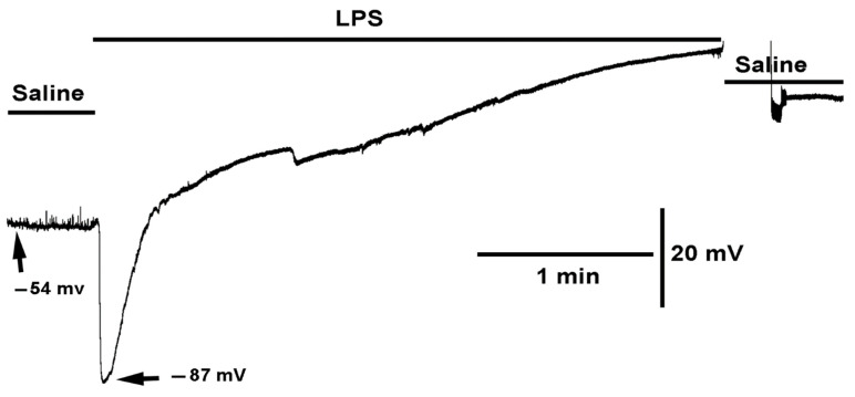
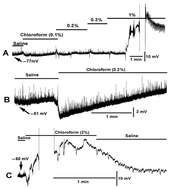
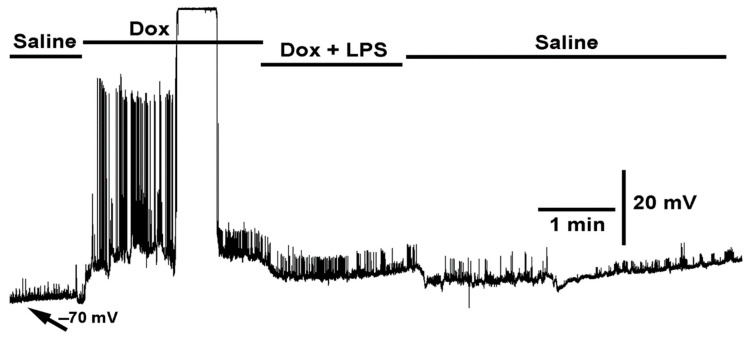
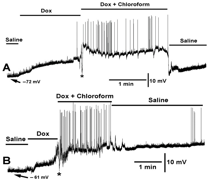
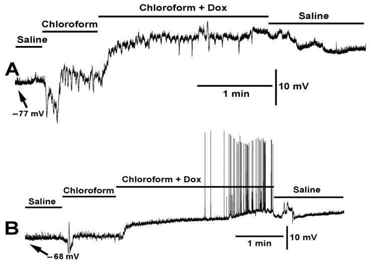
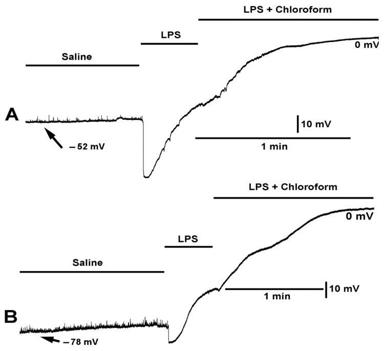
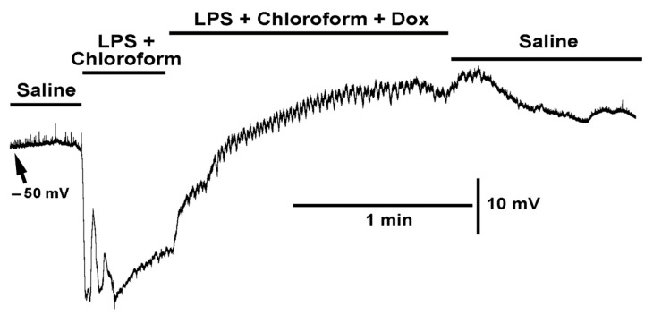
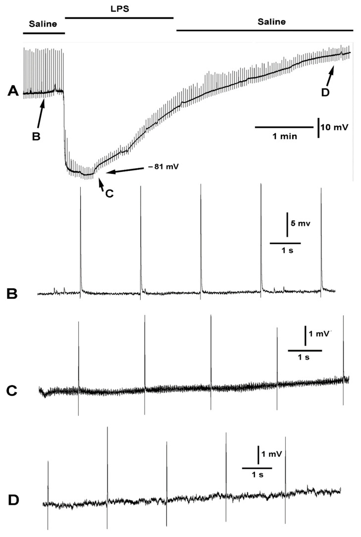
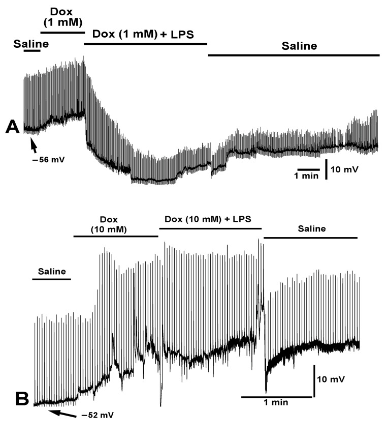
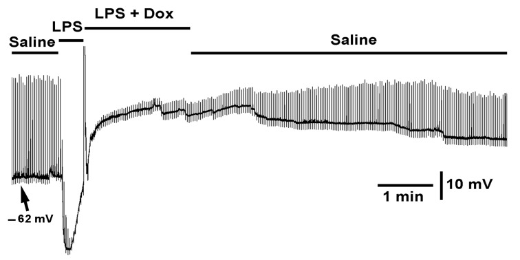
Bacterial septicemia is commonly induced by Gram-negative bacteria. The immune response is triggered in part by the secretion of bacterial endotoxin lipopolysaccharide (LPS). LPS induces the subsequent release of inflammatory cytokines which can result in pathological conditions. There is no known blocker to the receptors of LPS. The Drosophila larval muscle is an amendable model to rapidly screen various compounds that affect membrane potential and synaptic transmission such as LPS. LPS induces a rapid hyperpolarization in the body wall muscles and depolarization of motor neurons. These actions are blocked by the compound doxapram (10 mM), which is known to inhibit a subtype of the two-P-domain K+ channel (K2P channels). However, the K2P channel blocker PK-THPP had no effect on the Drosophila larval muscle at 1 and 10 mM. These channels are activated by chloroform, which also induces a rapid hyperpolarization of these muscles, but the channels are not blocked by doxapram. Likewise, chloroform does not block the depolarization induced by doxapram. LPS blocks the postsynaptic glutamate receptors on Drosophila muscle. Pre-exposure to doxapram reduces the LPS block of these ionotropic glutamate receptors. Given that the larval Drosophila body wall muscles are depolarized by doxapram and hyperpolarized by chloroform, they offer a model to begin pharmacological profiling of the K2P subtype channels with the potential of identifying blockers for the receptors to mitigate the actions of the Gram-negative endotoxin LPS.

Keywords: Drosophila; K2P channels; chloroform; doxapram; glutamate receptors; lipopolysaccharides; membrane potential.


Conflict of interest statement

The authors declare no conflict of interest.

Figures

Figure 1
Figure 1
The effect of LPS on membrane potential and synaptic transmission. LPS rapidly hyperpolarized the muscle as well as blocked the receptors to glutamate as the spontaneous quantal events (miniature excitatory junction potentials, mEJPs) were no longer observed even though there was an increased driving force. With prolonged exposure to LPS the membrane potential depolarizes to zero.
Figure 2
Figure 2
Dose dependent effect of chloroform on membrane potential and muscle contraction. (A) Chloroform in saline produced little effect at 0.1% concentration on hyperpolarizing the membrane potential. As the percent of chloroform increased a more pronounced rapid hyperpolarization (<1 s) would occur. Random hyperpolarization occurred during the 0.2% exposure. As the percent of chloroform reached 1% the muscle produced strong contractions, dislodging the intracellular electrode, and would continue to produce waves of contractions as shown at the end of the trace. (B) A saline with 0.2% chloroform produced a rapid hyperpolarization followed with a repolarization within 1 min. Note: The miniature excitatory junction potentials (mEJPs) are still present during the hyperpolarization. (C) At 2% chloroform, a rapid hyperpolarization occurred followed by strong muscle contraction making it difficult to maintain an intracellular recording. After reestablishing an intracellular recording in the same muscle, the repeated depolarization and hyperpolarization occurred due to muscle producing waves of contractions.
Figure 3
Figure 3
The effect of doxapram (10 mM) and the cocktail of doxapram (10 mM) and LPS on membrane potential and synaptic transmission. Exposure to doxapram (10 mM) rapidly depolarizes the membrane potential and evoked responses, from transected segmental nerves from the CNS. The motor neurons depolarize producing excitatory junction potentials (EJPs) of similar amplitude as those of electrical evoked nerve EJPs. The barraged of depolarizations dislodged the intracellular recordings. The bath exchange to doxapram and LPS produced small hyperpolarization while small EJPs and mEJPs are still present. Upon exchanging to fresh saline, free of doxapram and LPS, the membrane potential almost regained the initial resting membrane potential.
Figure 4
Figure 4
Two variations in the effect of doxapram (10 mM) and the cocktail of doxapram (10 mM) and chloroform (0.2%) are shown. (A,B) illustrate two different preparations of the same concentrations of compounds and mixtures. In both variations, doxapram depolarized the membrane potential and upon switching to the cocktail of doxapram (10 mM) and chloroform (0.2%), the membrane rapidly hyperpolarized (<1 s, see asterisks *) followed with depolarization and doxapram induced EJPs. Exchange to fresh saline recovered the membrane potential in (A) but doxapram had a lingering effect in some preparations as shown in (B).
Figure 5
Figure 5
Two examples of the effect on membrane potential by chloroform (0.2%) and chloroform (0.2%) combined with doxapram (10 mM) are shown (A,B). Two different preparations are shown with the same mixtures of compounds. Upon exposure to chloroform the membrane rapidly hyperpolarized followed by depolarization. In (A), chloroform produced a series of contractions. With the exposure to chloroform and doxapram the membrane depolarized even more. In (B), doxapram induced EJPs, while in A the effect of chloroform continues to produce waves of contraction.
Figure 6
Figure 6
The effect on the resting membrane potential by LPS and LPS combined with chloroform (0.2%). (A,B) Two different preparations are shown with the same mixtures of compounds. LPS produced a larger hyperpolarization in preparation shown in (A) than in the preparation shown in (B). Upon exposure of LPS combined with chloroform the membrane potential continued to depolarize to zero.
Figure 7
Figure 7
The effect on the resting membrane potential with exposure to LPS combined with chloroform (0.2%) and LPS combined with both chloroform (0.2%) and doxapram (10 mM). The combined LPS with chloroform still produced rapid hyperpolarization, but with some rapid depolarizations which is unlike LPS exposure by itself. LPS combined with both chloroform and doxapram rapidly depolarized the membrane and resulted in waves on contraction. The effects of the cocktail can partially be reversed by saline wash.
Figure 8
Figure 8
The effect of LPS on evoked synaptic transmission and resting membrane potential. The segmental nerve was stimulated at a rate of 0.5 Hz. (A) The overall effect on the membrane potential and the amplitude of the EJPs is shown. Note the membrane potential started to repolarize while still exposed to LPS. (B) The EJPs and mEJPs were observed in saline. (C) Exposure to LPS rapidly hyperpolarized the membrane and reduced the amplitude of the evoked EJPs and the mEJPs. The mEJPs can no longer be observed. Despite the increased driving force for increased amplitude of the EJPs and mEJPs they were greatly reduced in amplitude. (D) When the bath was exchanged to fresh saline, the membrane regains the initial value and the amplitude of the evoked EJPs increased.
Figure 9
Figure 9
The effect of doxapram (1 and 10 mM) and a cocktail of doxapram (1 and 10 mM) with LPS on membrane potential and evoked EJPs. The segmental nerve was stimulated at a rate of 0.5 Hz. (A,B) Two different preparations are shown with varied concentrations of doxapram. (A) A concentration of 1 mM doxapram resulted in depolarization of the membrane and the combination of doxapram (1 mM) with the same concentration (250 µg/mL) of LPS as used in B produced a hyperpolarization without the rapid depression in the amplitudes of the evoked EJPs. Upon exchanging the media for fresh saline, the amplitudes of the EJPs increased and the membrane potential trended towards regaining the initial value. (B) Doxapram at 10 mM produced a large depolarization and increased the amplitude of the evoked EJPs despite a reduced driving gradient due to the membrane being depolarized. The subsequent exchange of the media to doxapram (10 mM) and LPS (250 µg/mL) did not maintain a hyperpolarized state and the evoked as well as mEJPs were not greatly affected by the LPS. Upon exchanging the bath to fresh saline, the evoked EJPs reduced in amplitude but a high frequency of mEJPs remained.
Figure 10
Figure 10
The effect of LPS and the cocktail of LPS with doxapram on membrane potential and evoked EJPs. The segmental nerve was stimulated at a rate of 0.5 Hz. LPS rapidly hyperpolarized the membrane and depressed evoked synaptic transmission. The bath exchange to LPS and doxapram (10 mM) rapidly depolarized the membrane and does not fully relive the action of LPS in blocking synaptic transmission but allowed the synaptic response to increase in amplitude.

Similar articles

Cited by

References

    1. Osborn M.J., Rosen S.M., Rothfield L., Zeleznick L.D., Horecker B.L. Lipopolysaccharide of the gram-negative cell wall. Science. 1964;145:783–789. doi: 10.1126/science.145.3634.783. - DOI - PubMed
    1. Linhartová I., Bumba L., Mašín J., Basler M., Osička R., Kamanová J., Prochazkova K., Adkins I., Holubova J., Sadilkova L. RTX proteins: A highly diverse family secreted by a common mechanism. FEMS Microbiol. Rev. 2010;34:1076–1112. doi: 10.1111/j.1574-6976.2010.00231.x. - DOI - PMC - PubMed
    1. Da Silva Correia J., Soldau K., Christen U., Tobias P.S., Ulevitch R.J. Lipopolysaccharide is in close proximity to each of the proteins in its membrane receptor complex. transfer from CD14 to TLR4 and MD-2. J. Biol. Chem. 2001;276:21129–21135. doi: 10.1074/jbc.M009164200. - DOI - PubMed
    1. Wilson J.X., Young G.B. Progress in clinical neurosciences: Sepsis-associated encephalopathy: Evolving concepts. Can. J. Neurol. Sci. 2003;30:98–105. doi: 10.1017/S031716710005335X. - DOI - PubMed
    1. Friedrich O., Reid M.B., Van den Berghe G., Vanhorebeek I., Hermans G., Rich M.M., Larsson L. The sick and the weak: Neuropathies/myopathies in the critically ill. Physiol. Rev. 2015;95:1025–1109. doi: 10.1152/physrev.00028.2014. - DOI - PMC - PubMed

LinkOut - more resources