

Candida Administration in 5/6 Nephrectomized Mice Enhanced Fibrosis in Internal Organs: An Impact of Lipopolysaccharide and (1→3)-β-D-Glucan from Leaky Gut
- PMID: 36555628
- PMCID: PMC9784901
- DOI: 10.3390/ijms232415987
Candida Administration in 5/6 Nephrectomized Mice Enhanced Fibrosis in Internal Organs: An Impact of Lipopolysaccharide and (1→3)-β-D-Glucan from Leaky Gut
Abstract
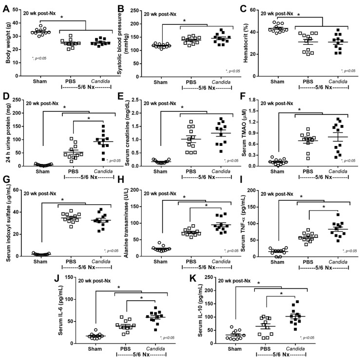
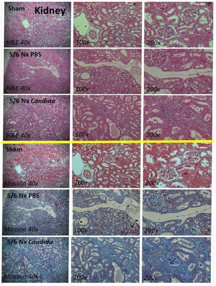
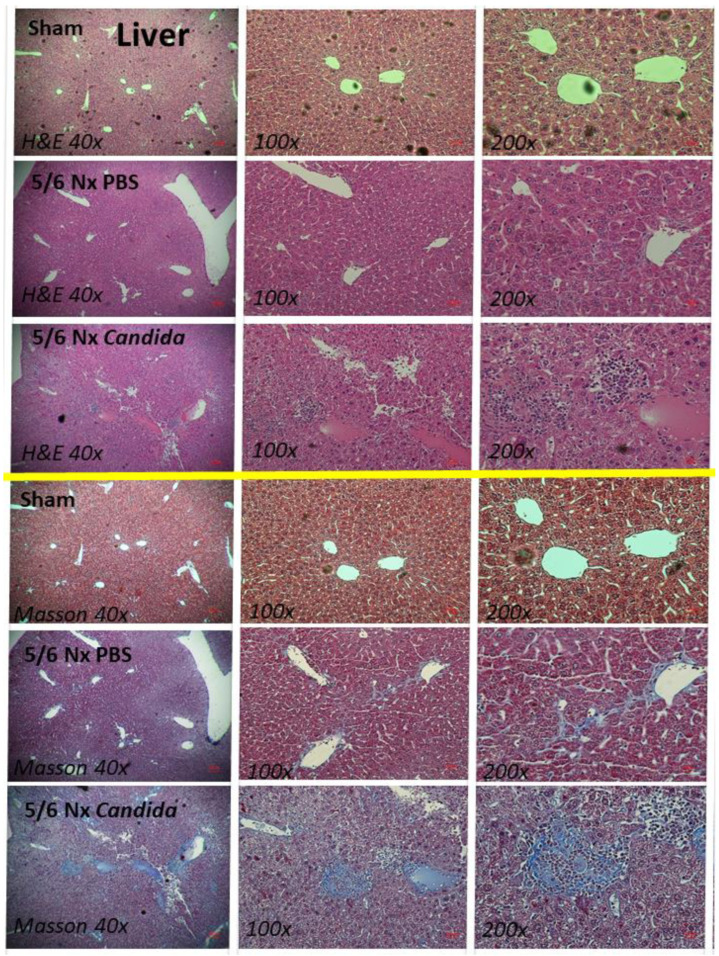
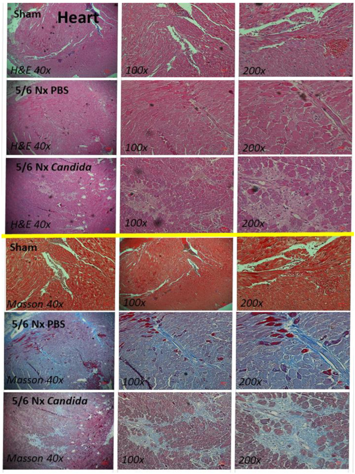
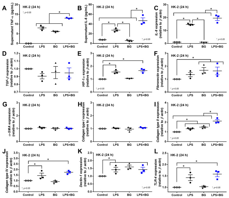
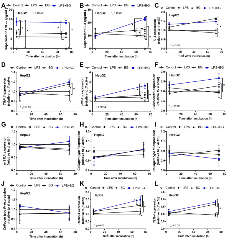
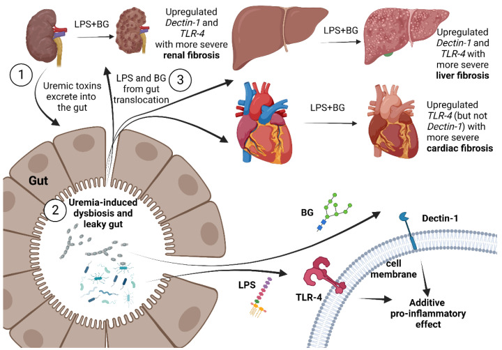
Uremic toxins and gut dysbiosis in advanced chronic kidney disease (CKD) can induce gut leakage, causing the translocation of gut microbial molecules into the systemic circulation. Lipopolysaccharide (LPS) and (1→3)-β-D-glucan (BG) are the major gut microbial molecules of Gram-negative bacteria and fungi, respectively, and can induce inflammation in several organs. Here, the fibrosis in the kidney, liver, and heart was investigated in oral C. albicans-administered 5/6 nephrectomized (Candida-5/6 Nx) mice. At 20 weeks post 5/6 Nx, Candida-5/6 Nx mice demonstrated increased 24 h proteinuria, liver enzymes, and serum cytokines (TNF-α, IL-6, and IL-10), but not weight loss, systolic blood pressure, hematocrit, serum creatinine, or gut-derived uremic toxins (TMAO and indoxyl sulfate), compared to in 5/6 Nx alone. The gut leakage in Candida-5/6 Nx was more severe, as indicated by FITC-dextran assay, endotoxemia, and serum BG. The areas of fibrosis from histopathology, along with the upregulated gene expression of Toll-like receptor 4 (TLR-4) and Dectin-1, the receptors for LPS and BG, respectively, were higher in the kidney, liver, and heart. In vitro, LPS combined with BG increased the supernatant IL-6 and TNF-α, upregulated the genes of pro-inflammation and pro-fibrotic processes, Dectin-1, and TLR-4 in renal tubular (HK-2) cells and hepatocytes (HepG2), when compared with LPS or BG alone. This supported the pro-inflammation-induced fibrosis and the possible LPS-BG additive effects on kidney and liver fibrosis. In conclusion, uremia-induced leaky gut causes the translocation of gut LPS and BG into circulation, which activates the pro-inflammatory and pro-fibrotic pathways, causing internal organ fibrosis. Our results support the crosstalk among several organs in CKD through a leaky gut.
Keywords: 5/6 nephrectomized mice; Candida; chronic kidney disease; fibrosis; gut leakage; gut-derived uremic toxins.
Conflict of interest statement
The authors declare no conflict of interest.
Figures

Similar articles
-
Uremia-Induced Gut Barrier Defect in 5/6 Nephrectomized Mice Is Worsened by Candida Administration through a Synergy of Uremic Toxin, Lipopolysaccharide, and (1➔3)-β-D-Glucan, but Is Attenuated by Lacticaseibacillus rhamnosus L34.Int J Mol Sci. 2022 Feb 24;23(5):2511. doi: 10.3390/ijms23052511. Int J Mol Sci. 2022. PMID: 35269654 Free PMC article.
-
Candida Administration Worsens Uremia-Induced Gut Leakage in Bilateral Nephrectomy Mice, an Impact of Gut Fungi and Organismal Molecules in Uremia.mSystems. 2021 Jan 12;6(1):e01187-20. doi: 10.1128/mSystems.01187-20. mSystems. 2021. PMID: 33436518 Free PMC article.
-
Candida Administration in Bilateral Nephrectomy Mice Elevates Serum (1→3)-β-D-glucan That Enhances Systemic Inflammation Through Energy Augmentation in Macrophages.Int J Mol Sci. 2021 May 10;22(9):5031. doi: 10.3390/ijms22095031. Int J Mol Sci. 2021. PMID: 34068595 Free PMC article.
-
Gut Leakage of Fungal-Derived Inflammatory Mediators: Part of a Gut-Liver-Kidney Axis in Bacterial Sepsis.Dig Dis Sci. 2019 Sep;64(9):2416-2428. doi: 10.1007/s10620-019-05581-y. Epub 2019 Mar 13. Dig Dis Sci. 2019. PMID: 30863955 Review.
-
Altered microbiome in chronic kidney disease: systemic effects of gut-derived uremic toxins.Clin Sci (Lond). 2018 Mar 9;132(5):509-522. doi: 10.1042/CS20171107. Print 2018 Mar 15. Clin Sci (Lond). 2018. PMID: 29523750 Review.
Cited by
-
Lacticaseibacillus rhamnosus attenuates uremic toxins in patients with nondialysis chronic kidney disease through the anti-inflammatory molecules.Sci Rep. 2025 Jul 31;15(1):27990. doi: 10.1038/s41598-025-12768-z. Sci Rep. 2025. PMID: 40744977 Free PMC article. Clinical Trial.
-
Gut microbiota regulates oxidative stress and inflammation: a double-edged sword in renal fibrosis.Cell Mol Life Sci. 2024 Dec 5;81(1):480. doi: 10.1007/s00018-024-05532-5. Cell Mol Life Sci. 2024. PMID: 39636415 Free PMC article. Review.
-
Kazachstania pintolopesii in Blood and Intestinal Wall of Macrophage-Depleted Mice with Cecal Ligation and Puncture, the Control of Fungi by Macrophages during Sepsis.J Fungi (Basel). 2023 Dec 4;9(12):1164. doi: 10.3390/jof9121164. J Fungi (Basel). 2023. PMID: 38132765 Free PMC article.
-
Lacticaseibacilli attenuated fecal dysbiosis and metabolome changes in Candida-administered bilateral nephrectomy mice.Front Immunol. 2023 Mar 9;14:1131447. doi: 10.3389/fimmu.2023.1131447. eCollection 2023. Front Immunol. 2023. PMID: 36969207 Free PMC article.
-
The evolving understanding of systemic mechanisms in organ-specific IgA nephropathy: a focus on gut-kidney crosstalk.Theranostics. 2025 Jan 1;15(2):656-681. doi: 10.7150/thno.104631. eCollection 2025. Theranostics. 2025. PMID: 39744688 Free PMC article. Review.
References
-
- Panpetch W., Kullapanich C., Dang C.P., Visitchanakun P., Saisorn W., Wongphoom J., Wannigama D.L., Thim-Uam A., Patarakul K., Somboonna N., et al. Candida Administration Worsens Uremia-Induced Gut Leakage in Bilateral Nephrectomy Mice, an Impact of Gut Fungi and Organismal Molecules in Uremia. Msystems. 2021;6:e01187-20. doi: 10.1128/mSystems.01187-20. - DOI - PMC - PubMed
MeSH terms
Substances
LinkOut - more resources
Full Text Sources
Medical
