Review
. 2022 Dec 15;23(24):15988.
doi: 10.3390/ijms232415988.

Molecular Regulation of Concomitant Lower Urinary Tract Symptoms and Erectile Dysfunction in Pelvic Ischemia

Affiliations
Review

Molecular Regulation of Concomitant Lower Urinary Tract Symptoms and Erectile Dysfunction in Pelvic Ischemia

Tufan Tarcan et al. Int J Mol Sci. .

Abstract

Aging correlates with greater incidence of lower urinary tract symptoms (LUTS) and erectile dysfunction (ED) in the male population where the pathophysiological link remains elusive. The incidence of LUTS and ED correlates with the prevalence of vascular risk factors, implying potential role of arterial disorders in concomitant development of the two conditions. Human studies have revealed lower bladder and prostate blood flow in patients with LUTS suggesting that the severity of LUTS and ED correlates with the severity of vascular disorders. A close link between increased prostatic vascular resistance and greater incidence of LUTS and ED has been documented. Experimental models of atherosclerosis-induced chronic pelvic ischemia (CPI) showed increased contractile reactivity of prostatic and bladder tissues, impairment of penile erectile tissue relaxation, and simultaneous development of detrusor overactivity and ED. In the bladder, short-term ischemia caused overactive contractions while prolonged ischemia provoked degenerative responses and led to underactivity. CPI compromised structural integrity of the bladder, prostatic, and penile erectile tissues. Downstream molecular mechanisms appear to involve cellular stress and survival signaling, receptor modifications, upregulation of cytokines, and impairment of the nitric oxide pathway in cavernosal tissue. These observations may suggest pelvic ischemia as an important contributing factor in LUTS-associated ED. The aim of this narrative review is to discuss the current evidence on CPI as a possible etiologic mechanism underlying LUTS-associated ED.

Keywords: atherosclerosis; bladder; erectile dysfunction; ischemia; lower urinary tract symptoms; oxidative stress; prostate.


Conflict of interest statement

The authors declare no conflict of interest.

Figures
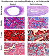
Figure 1
Figure 1
This figure shows that pelvic ischemia resulting from arterial atherosclerosis provokes simultaneous histological changes in the bladder, prostate, and penile erectile tissues. Iliac artery samples from animals with pelvic ischemia showed marked thickening of arterial intima and media layers and diminished arterial luminal size, suggesting atherosclerotic occlusive disease. Histologic assessment of tissue samples from the control bladders showed normal proportion of smooth muscle (red stain) and connective tissue (blue stain) with no indication of excessive collagen accumulation or fibrosis. Tissue samples from the ischemic bladder revealed dense bundles of collagen, suggesting diffuse fibrosis, loss of smooth muscle cells, and potential replacement of smooth muscle by connective tissue. Assessment of prostatic tissues showed swelling and thickening of the stroma, diminished smooth muscle density, accumulation of collagen, distorted glands with compressed epithelium lining, and the formation of large intraluminal spaces in the ischemic prostate tissues in comparison with control prostatic tissues. Consistent histological changes were evident in penile erectile tissue from animals with pelvic ischemia in comparison with controls. The erectile tissue smooth muscle content (stained red) was dramatically reduced, and connective tissue (stained blue) increased in the ischemic erectile tissue, suggesting loss of smooth muscle and diffuse fibrosis. The histology figures are shown at 40× magnification. These figures are taken from our previous publications [23,26,31].
Figure 2
Figure 2
Ultrastructural assessment of bladder, prostate, and penile erectile tissues by transmission electron microscopy revealed simultaneous widespread changes in the smooth muscle and nerve bundles of the ischemic samples versus controls. Deformed muscle fascicles, increased collagen deposition, swollen mitochondria with degraded granules, partial loss of mitochondrial membrane, and sporadic vacuolization were evident in both ischemic bladder and prostatic tissues. These changes were associated with cell shrinkage, condensation and fragmentation of the nucleus, folding and partial disruption of nuclear membrane, chromatin condensation, and increased cytoplasmic and nuclear ribosomes. Loss of endothelial integrity, increased caveolae, disrupted mitochondria with degraded granules, and sporadic vacuolization were found in the ischemic erectile tissues. Normal axon terminals packed with small vesicles, and normal ensheathed Schwann cells were found in the control bladder, prostate, and penile tissue samples. Degenerating axons with collapsed axonal and Schwann cell profile surrounded by dense connective tissue, sporadic vacuoles, and splitting of myelin sheath around both axon and Schwann cell were evident in the ischemic bladder, prostate and penile erectile tissues. M = mitochondria, N = nucleus, NM = nuclear membrane, C = caveolae, E = endothelium, V = vacuole. The bladder and prostate figures are reduced from 18,500x. The penile figures are reduced from 13,000x. These figures are taken from our previous publications [37,38,39].
Figure 3
Figure 3
This figure summarizes gene ontology analysis of the ncAA-containing proteins in bladder ischemia. The ncAA-containing proteins (R2 > 0.5, ratio > 2-fold, p < 0.05) in the ischemic bladder tissues were categorized using the PANTHER database. Changes in molecular function, biological process, cellular component, and protein class are presented in sections (a), (b), (c), and (d), respectively. These figures are taken from our previous publication [41].
Figure 4
Figure 4
Conceptual presentation of pelvic ischemia as a unifying mechanism of LUTS-associated ED. It is believed that bladder ischemia contributes to non-obstructive non-neurogenic detrusor overactivity. Prostate ischemia promotes prostatic smooth muscle contractions and leads to urinary outflow resistance independent of prostate size. Penile ischemia disrupts the underlying mechanism of erectile tissue relaxation and impairs erectile activity. These changes, cumulatively, lead to the development of LUTS-associated ED. NO = nitric oxide, cGMP = cyclic guanosine monophosphate, LUTS = lower urinary tract symptoms, ED = erectile dysfunction.

Similar articles

Cited by

References

    1. Diokno A.C., Brown M.B., Goldstein N.G., Herzog A.R. Urinary Flow Rates and Voiding Pressures in Elderly Men Living in a Community. J. Urol. 1994;151:1550–1553. doi: 10.1016/S0022-5347(17)35299-0. - DOI - PubMed
    1. Madersbacher S., Klingler H.C., Schatzl G., Stulnig T., Schmidbauer C.P., Marberger M. Age related urodynamic changes in patients with benign prostatic hyperplasia. J. Urol. 1996;156:1662–1667. doi: 10.1016/S0022-5347(01)65478-8. - DOI - PubMed
    1. Slayday R.E., Bell T.R., Lyons M.J., Toomey R., Vandiver R., Sliwinski M.J., Kremen W.S., Franz C.E. Erectile Function, Sexual Satisfaction, and Cognitive Decline in Men from Midlife to Older Adulthood. Gerontologist. 2022 doi: 10.1093/geront/gnac151. - DOI - PMC - PubMed
    1. Aleksandra R., Aleksandra S., Iwona R. Erectile Dysfunction in Relation to Metabolic Disorders and the Concentration of Sex Hormones in Aging Men. Int. J. Environ. Res. Public Health. 2022;19:7576. doi: 10.3390/ijerph19137576. - DOI - PMC - PubMed
    1. Feldman H.A., Goldstein I., Hatzichristou D.G., Krane R.J., McKinlay J.B. Impotence and Its Medical and Psychosocial Correlates: Results of the Massachusetts Male Aging Study. J. Urol. 1994;151:54–61. doi: 10.1016/S0022-5347(17)34871-1. - DOI - PubMed