

From Low-Grade Inflammation in Osteoarthritis to Neuropsychiatric Sequelae: A Narrative Review
- PMID: 36555670
- PMCID: PMC9784931
- DOI: 10.3390/ijms232416031
From Low-Grade Inflammation in Osteoarthritis to Neuropsychiatric Sequelae: A Narrative Review
Abstract
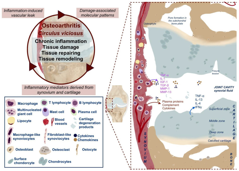
Nowadays, osteoarthritis (OA), a common, multifactorial musculoskeletal disease, is considered to have a low-grade inflammatory pathogenetic component. Lately, neuropsychiatric sequelae of the disease have gained recognition. However, a link between the peripheral inflammatory process of OA and the development of neuropsychiatric pathology is not completely understood. In this review, we provide a narrative that explores the development of neuropsychiatric disease in the presence of chronic peripheral low-grade inflammation with a focus on its signaling to the brain. We describe the development of a pro-inflammatory environment in the OA-affected joint. We discuss inflammation-signaling pathways that link the affected joint to the central nervous system, mainly using primary sensory afferents and blood circulation via circumventricular organs and cerebral endothelium. The review describes molecular and cellular changes in the brain, recognized in the presence of chronic peripheral inflammation. In addition, changes in the volume of gray matter and alterations of connectivity important for the assessment of the efficacy of treatment in OA are discussed in the given review. Finally, the narrative considers the importance of the use of neuropsychiatric diagnostic tools for a disease with an inflammatory component in the clinical setting.
Keywords: central nervous system; cytokines; low-grade inflammation; neuropsychiatric disease; osteoarthritis; signaling.
Conflict of interest statement
The authors declare no conflict of interest.
Figures


Similar articles
-
Metabolic triggered inflammation in osteoarthritis.Osteoarthritis Cartilage. 2015 Jan;23(1):22-30. doi: 10.1016/j.joca.2014.10.002. Epub 2014 Oct 15. Osteoarthritis Cartilage. 2015. PMID: 25452156 Review.
-
Osteoarthritis, cerebrovascular dysfunction and the common denominator of inflammation: a narrative review.Osteoarthritis Cartilage. 2018 Apr;26(4):462-470. doi: 10.1016/j.joca.2018.01.011. Epub 2018 Feb 2. Osteoarthritis Cartilage. 2018. PMID: 29406252 Review.
-
[AGING AND OSTEOARTHRITIS. CHRONIC NONSPECIFIC INFLAMMATION AS A LINK BETWEEN AGING AND OSTEOARTHRITIS (REVIEW)].Adv Gerontol. 2015;28(2):274-83. Adv Gerontol. 2015. PMID: 26856088 Review. Russian.
-
Role of the Sympathetic Nervous System in Mild Chronic Inflammatory Diseases: Focus on Osteoarthritis.Neuroimmunomodulation. 2023;30(1):143-166. doi: 10.1159/000531798. Epub 2023 Jul 10. Neuroimmunomodulation. 2023. PMID: 37429263 Free PMC article. Review.
-
[Inflammation and osteoarthritis-related pain].Schmerz. 2019 Feb;33(1):4-12. doi: 10.1007/s00482-018-0346-y. Schmerz. 2019. PMID: 30560495 Review. German.
Cited by
-
PSD95 as a New Potential Therapeutic Target of Osteoarthritis: A Study of the Identification of Hub Genes through Self-Contrast Model.Int J Mol Sci. 2023 Sep 28;24(19):14682. doi: 10.3390/ijms241914682. Int J Mol Sci. 2023. PMID: 37834131 Free PMC article.
-
Obesity, Metabolic Syndrome, and Osteoarthritis Require Integrative Understanding and Management.Biomedicines. 2024 Jun 6;12(6):1262. doi: 10.3390/biomedicines12061262. Biomedicines. 2024. PMID: 38927469 Free PMC article. Review.
-
Identification of Novel Diagnostic Biomarkers and Classification Patterns for Osteoarthritis by Analyzing a Specific Set of Genes Related to Inflammation.Inflammation. 2023 Dec;46(6):2193-2208. doi: 10.1007/s10753-023-01871-w. Epub 2023 Jul 18. Inflammation. 2023. PMID: 37462886
-
Estimating the impact of metabolic syndrome on low back pain and the joint effects of metabolic syndrome and depressive symptoms on low back pain: insights from the China Health and Retirement Longitudinal Study.BMC Public Health. 2024 Aug 30;24(1):2359. doi: 10.1186/s12889-024-19851-6. BMC Public Health. 2024. PMID: 39215306 Free PMC article.
References
Publication types
MeSH terms
Grants and funding
LinkOut - more resources
Full Text Sources
Medical
