. 2022 Dec 16;23(24):16087.
doi: 10.3390/ijms232416087.

iPSC-Derived Macrophages: The Differentiation Protocol Affects Cell Immune Characteristics and Differentiation Trajectories

Affiliations

iPSC-Derived Macrophages: The Differentiation Protocol Affects Cell Immune Characteristics and Differentiation Trajectories

Anna Klepikova et al. Int J Mol Sci. .

Abstract

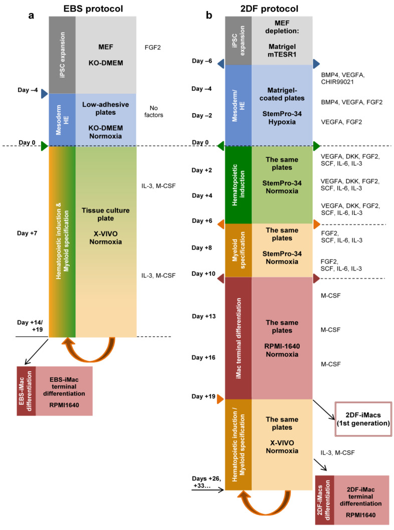
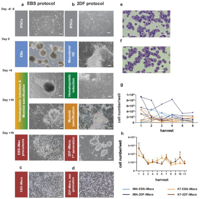
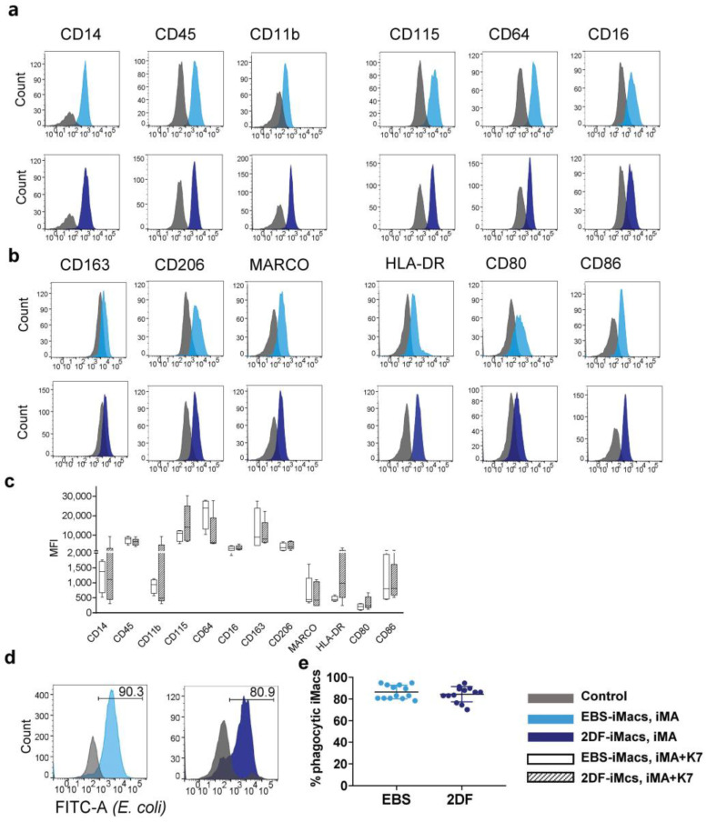
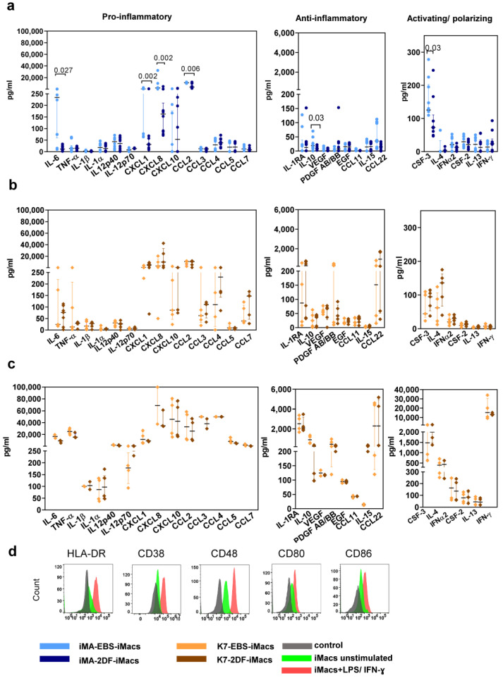
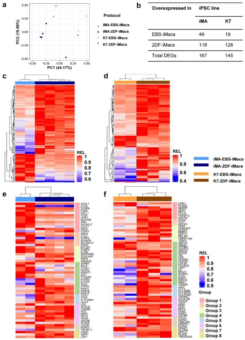
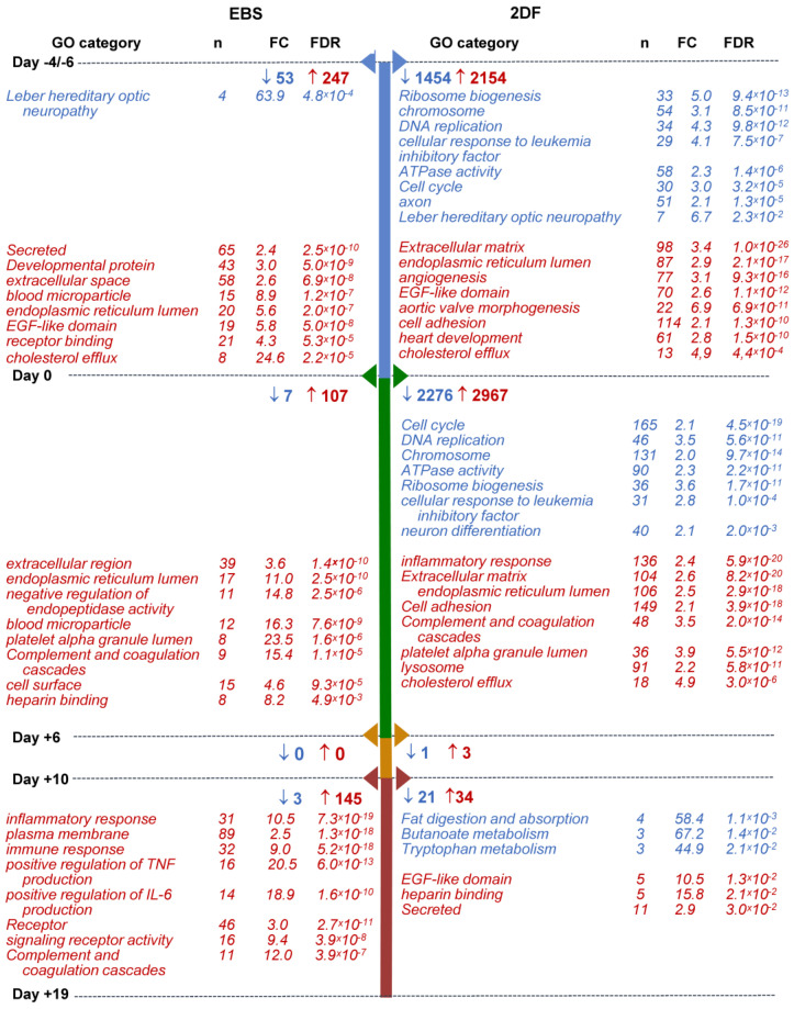
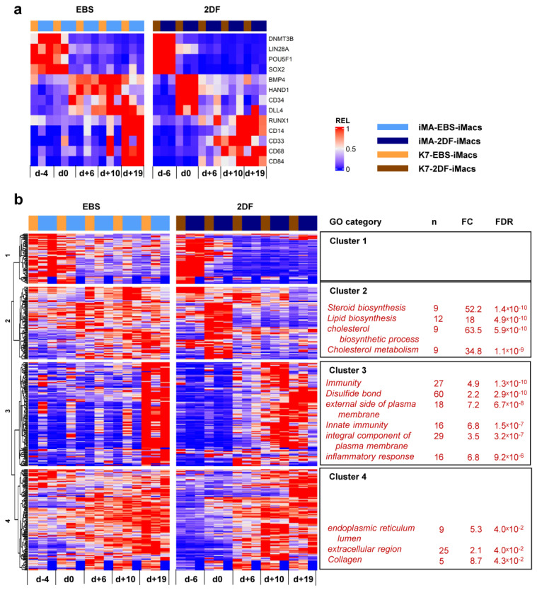
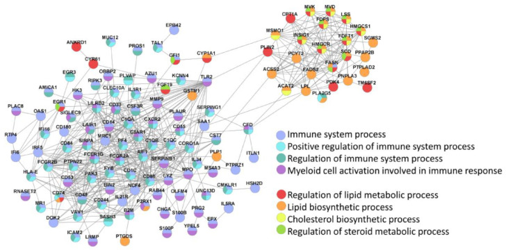
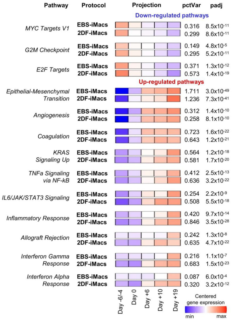
The generation of human macrophages from induced pluripotent stem cells (iMacs) is a rapidly developing approach used to create disease models, screen drugs, study macrophage-pathogen interactions and develop macrophage-based cell therapy. To generate iMacs, different types of protocols have been suggested, all thought to result in the generation of similar iMac populations. However, direct comparison of iMacs generated using different protocols has not been performed. We have compared the productivity, the differentiation trajectories and the characteristics of iMacs generated using two widely used protocols: one based on the formation of embryoid bodies and the induction of myeloid differentiation by only two cytokines, interleukin-3 and macrophage colony-stimulating factor, and the other utilizing multiple exogenous factors for iMac generation. We report inter-protocol differences in the following: (i) protocol productivity; (ii) dynamic changes in the expression of genes related to inflammation and lipid homeostasis following iMac differentiation and (iii) the transcriptomic profiles of terminally differentiated iMacs, including the expression of genes involved in inflammatory response, antigen presentation and lipid homeostasis. The results document the dependence of fine iMac characteristics on the type of differentiation protocol, which is important for further development of the field, including the development of iMac-based cell therapy.

Keywords: antigen presentation; induced pluripotent stem cells; inflammatory response; lipid homeostasis; macrophage differentiation; macrophages derived from induced pluripotent stem cells.


Conflict of interest statement

The authors declare no conflict of interest.

Figures

Figure 1
Figure 1
Schematic representation of iMac differentiation procedures using embryoid-body-dependent spontaneous (EBS) and embryoid-body-independent exogenous factor-dependent (2DF) differentiation protocols. (a) EBS protocol. induced pluripotent stem cells (iPSCs) were expanded on mouse embryonic fibroblast feeder cells (MEFs) and collected and cultured in low-adherent conditions in the absence of exogenous differentiation factors (day −4 to day 0) to induce embryoid body (EB) formation. At day 0, EBs were collected and transferred to culture tissue plates, where they were cultured in the presence of IL-3 and macrophage colony-stimulating factor (M-CSF) to induce hematopoietic specification and myeloid differentiation. When floating iMac precursors appeared in the culture (day +14 to day +19), they were collected and differentiated into EBS-iMacs in the presence of M-CSF. The remaining cultures were restimulated with IL-3/M-CSF to continue the generation of EBS-iMac precursors followed by their terminal differentiation into EBS-iMacs. (b) The 2DF protocol. To induce mesoderm/HE, MEF-depleted iPSCs were cultured in tissue culture plates coated with Matrigel in the presence of indicated exogenous factors (day −6 to day 0). On day 0, the composition of exogenous factors was changed in a way to induce first hematopoietic specification (day 0 to day +6) and next myeloid differentiation (day +6 to day +10). BMP4, bone morphogenetic protein 4; DKK, Dickkopf-related protein 1; FGF2, basic fibroblast growth factor; Flt3L, Fms-related receptor tyrosine kinase 3 ligand; SCF, stem cell factor; TPO, thrombopoietin; VEGFA, vascular endothelial growth factor A.
Figure 2
Figure 2
Representative morphology of differentiating cells, resulting iMac populations and harvest counts of EBS- and 2DF-iMacs. (a,b) Representative light microscopy of cell cultures at the indicated stages of iMac differentiation. (a) EBS protocol, (b) 2DF protocol. (c,d) Representative light microscopy of EBS- (c) and 2DF (d) iMacs. Phase contrast. (e,f) Giemsa staining of EBS-iMac (e) and 2DF-iMac (f) cytospins. (af) Scale bars represent 100 µm; shown are representative results obtained during the differentiation of iPSC line K7; similar data were obtained for iPSC line iMA. (g) Weekly yields of EBS- and 2DF-iMacs derived from iPSC line iMA (blue) and K7 (orange). Light colors, EBS-iMacs; dark colors, 2DF-iMacs. Most experiments were terminated at week 6. (h) A comparison of the duration of EBS- and 2DF-iMac generation summarized data obtained in 5 independent differentiation experiments for each type of protocol (iPSC line K7).
Figure 3
Figure 3
EBS- and 2DF-iMacs display similar phenotype and phagocytic activity. (ad) EBS- and 2DF-iMacs were collected at different differentiation time points and stained with antibodies or analyzed in PhagotestTM (d,e). (a) iMac expression of markers specific for macrophages/myeloid cells. (b) iMac expression of markers associated with macrophage polarization and activation. Shown are the results of one experiment obtained in iMA-derived EBS-iMacs and 2DF-iMacs. The results are representative of 7 independent experiments performed in iMA-derived (n = 3) and K7-derived (n = 4) EBS- and 2DF-iMacs; EBS-iMacs and 2DF-iMacs were differentiated and analyzed simultaneously; in each experiment, 3000 to 10,000 CD14+ cells were collected; for histograms, data were downsampled to 3000 events. (c) Mean fluorescence intensity (MFI) of the expression of analyzed markers on iMA- and K7-derived EBS- and 2DF-iMacs: summarized data obtained in 6 independent experiments (iMA, n = 3; K7, n = 3). (d) The comparison of the phagocytic activity of EBS- and 2DF-iMacs in PhagotestTM: representative histograms. (e) The comparison of the phagocytic activity of EBS- and 2DF-iMacs in PhagotestTM: summarized data (13 independent experiments; shown are percentages of phagocytic cells after gating on CD14+ cells, mean ± SD). Analyses were performed using iMac harvests 2–17 and gave similar results. In each experiment, at least 5000 CD14+ cells were collected and analyzed.
Figure 4
Figure 4
EBS- and 2DF-iMacs are responsive to inflammatory stimuli. EBS- and 2DF-iMacs were left unstimulated or were stimulated with LPS (100 ng/mL) and IFN-γ (20 ng/mL); 24 h later, the supernatants were collected. The cells were stained with antibodies specific to HLA-DR, CD38, CD48, CD80 and CD86. (a,b) The baseline secretion of indicated cytokines by EBS- and 2DF-iMacs (medians and interquartile ranges). (a) iMA-derived iMacs; (b) K7-derived iMacs. (c) The secretion of indicated cytokines by K7-derived iMacs stimulated with LPS/IFN-γ. Similar results were obtained using iMA-derived iMacs. The supernatants were collected from iMacs generated in 3 independent iMA and 3 independent K7 differentiation experiments. In each differentiation experiment, the supernatants were collected from 1–2 iMac harvests; all supernatants were analyzed simultaneously. (d) Surface expression of HLA-DR, CD38, CD48, CD80 and CD86 by EBS- and 2DF-iMacs (representative data of 3 independent experiments obtained in K7-derived iMacs).
Figure 5
Figure 5
EBS- and 2DF-iMacs differ by fine transcriptomic characteristics. EBS- and 2DF-iMacs were differentiated in parallel in 4 (iMA) and 3 (K7) independent differentiation experiments. RNA was isolated from the resulting CD14+ sorted iMacs (provided they were formed on time) and sequenced (two batches of iMA-EBS-iMacs and one batch of K7-EBS-iMacs were generated late and were excluded from the analysis). (a) Principal component (PC) analysis showing the separation of iMacs based on the source iPSC line (PC1) and the type of differentiation protocol (PC2). (b) Numbers of differentially expressed genes (DEGs) between iMA- and K7-derived EBS-iMacs and 2DF-iMacs. (c,d) Heatmaps depicting the expression of DEGs in iMA- (c) and K7- (d) derived EBS-iMacs and 2DF-iMacs. Expression levels were normalized on the maximal value separately for (i) each gene and (ii) EBS and 2DF differentiation experiments. (e,f) Heatmaps depicting the expression of selected DEGs between EBS- and 2DF-iMacs. (e) iMA-derived iMacs; (f) K7-derived iMacs. DEGs were categorized into 8 functional groups based on gene role in macrophage functionality taken from available published sources. Group 1, genes induced by M1/inflammatory stimuli and involved in pro-inflammatory response; group 2, genes induced by M1/inflammatory stimuli for which both pro- and anti-inflammatory effects have been reported; group 3, genes induced by M1/inflammatory stimuli and involved in the negative regulation of inflammation; group 4, genes associated with M2/TAM macrophages and anti-inflammatory activity; group 5, genes implicated in antigen presentation, endosome functioning and costimulation; group 6, genes involved in lipid homeostasis and foam macrophage formation; group 7, genes implicated in osteoclastogenesis; group 8, genes implicated in phagocytosis and antibacterial response. For detailed description of genes, see Tables S2.1–S2.3.
Figure 6
Figure 6
Selected categories of genes down- and up-regulated following iPSC to iMac differentiation using EBS (left) and 2DF (right) protocols (intra-protocol comparison). Three parallel EBS and 2DF differentiation experiments were performed using iMA (n = 2) and K7 (n = 1) iPSCs. RNA and 2DF differentiation experiments were performed using iMA (n = 2) and K7 (n = 1) iPSCs. RNA was isolated from iPSCs and differentiating cells on days d−4/−6, d0, d+6, d+10 and d+19; genes that significantly changed their expression in all three differentiation experiments at each differentiation stage within a given protocol (“intra-protocol pairwise DEGs”) were determined using DESeq2. GO terms and other categories shown in the figure were selected based on the following criteria: (i) are referred to in Gene Ontology (GO), Kyoto Encyclopedia of Genes and Genomes (KEGG) or UniProtKB Keywords (UP-KW); (ii) the false discovery rate (FDR) is <0.05; (iii) fold change (FC) is >2; (iv) if several structurally or functionally similar GO terms/other categories met criteria i-iii, only one of them (having the lowest FDR) was selected. Blue, GOs and other categories down-regulated at the indicated differentiation stages; red, GOs and other categories up-regulated at the indicated differentiation stages. For the entire list of GO terms and other categories, see Tables S4.1–S4.4.
Figure 7
Figure 7
Dynamic transcriptomic profiles of cells undergoing differentiation using EBS and 2DF protocols (inter-protocol differences). The cells were differentiated, and RNA was isolated as described in Figure 6. (a) Dynamic changes in the expression of selected genes associated with (i) cell pluripotency (DNMT3B, LIN28A, POU5F1 and SOX2); (ii) mesoderm (BMP4 and HAND1); (iii) hemogenic endothelium and hematopoietic precursors (CD34, DLL4 and RUNX1); and (iv) myeloid cells/macrophages (CD14, CD33, CD68 and CD84). (b) Inter-protocol DEGs identified by applying DESeq2 to all RNA-seq data obtained in three differentiation experiments in both protocols at all differentiation time points. For each cluster, selected GO terms are listed.
Figure 8
Figure 8
STRING identifies two main clusters of inter-protocol differentially expressed genes that are associated with immune response and lipid homeostasis. All inter-protocol DEGs (Figure 7b) were uploaded to STRING. The input options included a confidence cutoff of 0.4 and maximum additional interactions of 0. The GO Biological Processes with FDR < 4.0 × 10⁻⁶ are shown.
Figure 9
Figure 9
Pathway-level analysis highlights dynamic expression changes in cells differentiating in EBS and 2DF protocols. RNA-seq data obtained in two independent differentiation experiments were analyzed using the gene set co-regulation analysis (GESECA) which computes percent of explained variance for each of the gene sets (pathways) from human Hallmark collection of Molecular Signatures Database (each differentiation experiment included parallel differentiation of iPSC line iMA using EBS and 2DF protocols). Significant pathways associated with cell proliferative and immunological activity and differentiation processes are shown (inter-protocol pathways). Projection illustrates centered gene expression dynamics with colors scaled for each protocol individually from minimal to maximal value of the analyzed expression matrix; pctVar—percent of variance in the data that the corresponding pathway explains; padj—adjusted p-value. Similar results were obtained for the differentiation of iPSC line K7 (one differentiation experiment, the results are presented in Figure S1).

References

    1. Panicker L.M., Miller D., Park T.S., Pate B., Azevedo J.L., Awad O., Masood M.A., Veenstra T.D., Goldin E., Stubblefield B.K., et al. Induced pluripotent stem cell model recapitulates pathologic hallmarks of Gaucher disease. Proc. Natl. Acad. Sci. USA. 2012;109:18054–18059. doi: 10.1073/pnas.1207889109. - DOI - PMC - PubMed
    1. Brault J., Goutagny E., Telugu N., Shao K., Baquié M., Satre V., Coutton C., Grunwald D., Brion J.P., Barlogis V., et al. Optimized generation of functional neutrophils and macrophages from patient-specific induced pluripotent stem cells: Ex vivo models of X(0)-linked, AR22(0)- and AR47(0)- chronic granulomatous diseases. BioResearch. 2014;3:311–326. doi: 10.1089/biores.2014.0045. - DOI - PMC - PubMed
    1. Zhang H., Xue C., Shah R., Bermingham K., Hinkle C.C., Li W., Rodrigues A., Tabita-Martinez J., Millar J.S., Cuchel M., et al. Functional analysis and transcriptomic profiling of iPSC-derived macrophages and their application in modeling Mendelian disease. Circ. Res. 2015;117:17–28. doi: 10.1161/CIRCRESAHA.117.305860. - DOI - PMC - PubMed
    1. Klatt D., Cheng E., Philipp F., Selich A., Dahlke J., Schmidt R.E., Schott J.W., Büning H., Hoffmann D., Thrasher A.J., et al. Targeted repair of p47-CGD in iPSCs by CRISPR/Cas9: Functional correction without cleavage in the highly homologous pseudogenes. Stem Cell Rep. 2019;13:590–598. doi: 10.1016/j.stemcr.2019.08.008. - DOI - PMC - PubMed
    1. Zhang H., Reilly M.P. Human induced pluripotent stem cell-derived macrophages for unraveling human macrophage biology. Arterioscler. Throm. Vasc. Biol. 2017;37:2000–2006. doi: 10.1161/ATVBAHA.117.309195. - DOI - PMC - PubMed