

LAIR1, an ITIM-Containing Receptor Involved in Immune Disorders and in Hematological Neoplasms
- PMID: 36555775
- PMCID: PMC9788452
- DOI: 10.3390/ijms232416136
LAIR1, an ITIM-Containing Receptor Involved in Immune Disorders and in Hematological Neoplasms
Abstract
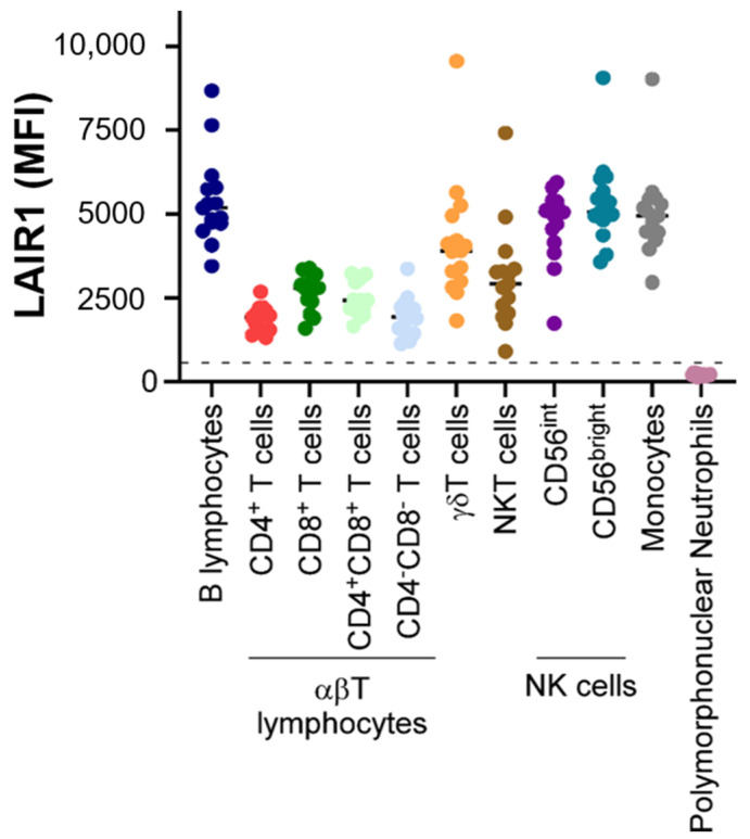
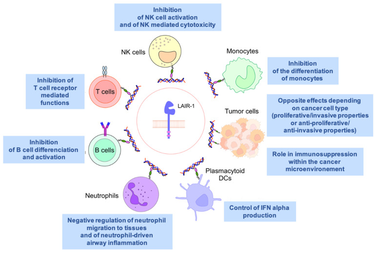
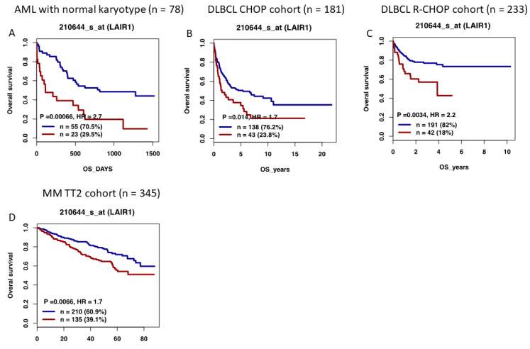
Leukocyte-associated immunoglobulin (Ig)-like receptor 1 (LAIR1, CD305) belongs to the family of immune-inhibitory receptors and is widely expressed on hematopoietic mature cells, particularly on immune cells. Four different types of ligands of LAIR1 have been described, including collagens, suggesting a potential immune-regulatory function on the extracellular matrix. By modulating cytokine secretion and cellular functions, LAIR1 displays distinct patterns of expression among NK cell and T/B lymphocyte subsets during their differentiation and cellular activation and plays a major negative immunoregulatory role. Beyond its implications in physiology, the activity of LAIR1 can be inappropriately involved in various autoimmune or inflammatory disorders and has been implicated in cancer physiopathology, including hematological neoplasms. Its action as an inhibitory receptor can result in the dysregulation of immune cellular responses and in immune escape within the tumor microenvironment. Furthermore, when expressed by tumor cells, LAIR1 can modulate their proliferation or invasion properties, with contradictory pro- or anti-tumoral effects depending on tumor type. In this review, we will focus on its role in normal physiological conditions, as well as during pathological situations, including hematological malignancies. We will also discuss potential therapeutic strategies targeting LAIR1 for the treatment of various autoimmune diseases and cancer settings.
Keywords: LAIR1; autoimmunity; collagen; hematological neoplasms; immunoregulatory; inflammation; inhibitory receptor.
Conflict of interest statement
The authors declare no conflict of interest. The funders had no role in the design of the study; in the collection, analyses, or interpretation of data; in the writing of the manuscript; or in the decision to publish the results.
Figures

Similar articles
-
Leukocyte-Associated Ig-like Receptor 1 Inhibits Th1 Responses but Is Required for Natural and Induced Monocyte-Dependent Th17 Responses.J Immunol. 2018 Jul 15;201(2):772-781. doi: 10.4049/jimmunol.1701753. Epub 2018 Jun 8. J Immunol. 2018. PMID: 29884698 Free PMC article.
-
Inhibitory pattern recognition receptors: lessons from LAIR1.Nat Rev Immunol. 2025 May 27. doi: 10.1038/s41577-025-01181-2. Online ahead of print. Nat Rev Immunol. 2025. PMID: 40425821 Review.
-
Blocking LAIR1 signaling in immune cells inhibits tumor development.Front Immunol. 2022 Sep 21;13:996026. doi: 10.3389/fimmu.2022.996026. eCollection 2022. Front Immunol. 2022. PMID: 36211388 Free PMC article.
-
A perspective on LILRBs and LAIR1 as immune checkpoint targets for cancer treatment.Biochem Biophys Res Commun. 2022 Dec 10;633:64-67. doi: 10.1016/j.bbrc.2022.09.019. Biochem Biophys Res Commun. 2022. PMID: 36344166 Review.
-
Antitumor Activity of a Novel LAIR1 Antagonist in Combination with Anti-PD1 to Treat Collagen-Rich Solid Tumors.Mol Cancer Ther. 2024 Aug 1;23(8):1144-1158. doi: 10.1158/1535-7163.MCT-23-0866. Mol Cancer Ther. 2024. PMID: 38648067 Free PMC article.
Cited by
-
Immune inhibitory receptor agonist therapeutics.Front Immunol. 2025 Mar 26;16:1566869. doi: 10.3389/fimmu.2025.1566869. eCollection 2025. Front Immunol. 2025. PMID: 40207220 Free PMC article. Review.
-
Dual-inhibitory domain iCARs improve the efficiency of the AND-NOT gate CAR T strategy.Proc Natl Acad Sci U S A. 2023 Nov 21;120(47):e2312374120. doi: 10.1073/pnas.2312374120. Epub 2023 Nov 14. Proc Natl Acad Sci U S A. 2023. PMID: 37963244 Free PMC article.
-
Collagen remodeling-mediated signaling pathways and their impact on tumor therapy.J Biol Chem. 2025 Mar;301(3):108330. doi: 10.1016/j.jbc.2025.108330. Epub 2025 Feb 19. J Biol Chem. 2025. PMID: 39984051 Free PMC article. Review.
-
Single cell RNA-seq reveals cellular and transcriptional heterogeneity in the splenic CD11b+Ly6Chigh monocyte population expanded in sepsis-surviving mice.Mol Med. 2024 Nov 6;30(1):202. doi: 10.1186/s10020-024-00970-0. Mol Med. 2024. PMID: 39506629 Free PMC article.
-
Sex effects on DNA methylation affect discovery in epigenome-wide association study of schizophrenia.Mol Psychiatry. 2024 Aug;29(8):2467-2477. doi: 10.1038/s41380-024-02513-9. Epub 2024 Mar 19. Mol Psychiatry. 2024. PMID: 38503926 Free PMC article.
References
-
- Verbrugge A., Ruiter Td T., Clevers H., Meyaard L. Differential contribution of the immunoreceptor tyrosine-based inhibitory motifs of human leukocyte-associated Ig-like receptor-1 to inhibitory function and phosphatase recruitment. Int. Immunol. 2003;15:1349–1358. doi: 10.1093/intimm/dxg134. - DOI - PubMed
-
- Lebbink R.J., van den Berg M.C., de Ruiter T., Raynal N., van Roon J.A., Lenting P.J., Jin B., Meyaard L. The soluble leukocyte-associated Ig-like receptor (LAIR)-2 antagonizes the collagen/LAIR1 inhibitory immune interaction. J. Immunol. 2008;180:1662–1669. doi: 10.4049/jimmunol.180.3.1662. - DOI - PubMed
Publication types
MeSH terms
Grants and funding
LinkOut - more resources
Full Text Sources
Medical
