. 2022 Dec 17;23(24):16137.
doi: 10.3390/ijms232416137.

Targeting TLR2/Rac1/cdc42/JNK Pathway to Reveal That Ruxolitinib Promotes Thrombocytopoiesis

Affiliations

Targeting TLR2/Rac1/cdc42/JNK Pathway to Reveal That Ruxolitinib Promotes Thrombocytopoiesis

Shuo Yang et al. Int J Mol Sci. .

Abstract

Background: Thrombocytopenia has long been considered an important complication of chemotherapy and radiotherapy, which severely limits the effectiveness of cancer treatment and the overall survival of patients. However, clinical treatment options are extremely limited so far. Ruxolitinib is a potential candidate.

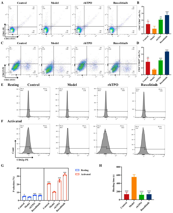
Methods: The impact of ruxolitinib on the differentiation and maturation of K562 and Meg-01 cells megakaryocytes (MKs) was examined by flow cytometry, Giemsa and Phalloidin staining. A mouse model of radiation-injured thrombocytopenia (RIT) was employed to evaluate the action of ruxolitinib on thrombocytopoiesis. Network pharmacology, molecular docking, drug affinity responsive target stability assay (DARTS), RNA sequencing, protein blotting and immunofluorescence analysis were applied to explore the targets and mechanisms of action of ruxolitinib.

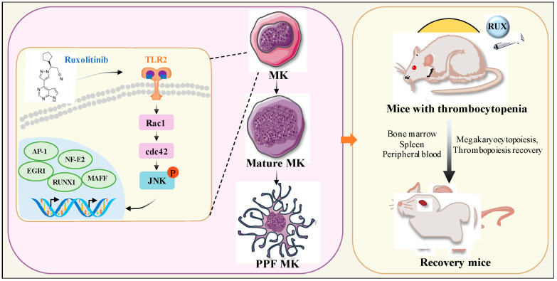
Results: Ruxolitinib can stimulate MK differentiation and maturation in a dose-dependent manner and accelerates recovery of MKs and thrombocytopoiesis in RIT mice. Biological targeting analysis showed that ruxolitinib binds directly to Toll Like Receptor 2 (TLR2) to activate Rac1/cdc42/JNK, and this action was shown to be blocked by C29, a specific inhibitor of TLR2.

Conclusions: Ruxolitinib was first identified to facilitate MK differentiation and thrombocytopoiesis, which may alleviate RIT. The potential mechanism of ruxolitinib was to promote MK differentiation via activating the Rac1/cdc42/JNK pathway through binding to TLR2.

Keywords: MK; TLR2/Rac1/cdc42/JNK; radiation; rusolitinib; thrombocytopoiesis.


Conflict of interest statement

The authors declare that the research was conducted in the absence of any commercial or financial relationships that could be construed as a potential conflict of interest.

Figures

Figure 1
Figure 1
Safe concentration of ruxolitinib for treatment of K562 and Meg-01. (A, B) The effect of Ruxolitinib intervened on MKs proliferation. Different time points and concentrations on the proliferation rate (%) of megaryocytes. Results of the CCK-8 assay for K562 and Meg-01 cells proliferation; (C, D) The LDH release of ruxolitinib intervened K562 and Meg01 cells at different time points; (E, F) Representative images of K562 and Meg-01 cells treated with various concentrations of Ruxolitinib (5, 10, and 20 μM) for 5 days. The positive control is PMA (2.5 nM). n = 3, * p < 0.05, ** p < 0.01, *** p < 0.001, vs. control.
Figure 2
Figure 2
Ruxolitinib induces dramatic morphological changes and differentiations. (A,B) The percentage of CD41+/CD42b+ complexes surface expression on K562 and Meg-01 cells by ruxolitinib (5, 10 and 20 μM) or PMA (2.5 nM) for 5 days and analyzed by flow cytometry; (C) Giemsa staining of K562 and Meg-01 cells treated with ruxolitinib (5, 10, and 20 μM). Magnification: 400×, Scale bar: 100 μm; (D) Phalloidin-labeled cytospin in K562 and Meg-01 cells on day 5 under a fluorescence Microscope (excitation wavelength: 560 nm for Phalloidin, 405 nm for DAPI). Magnification: 400×, Scale bar: 50 μm. n = 3, mean ± SD. Statistics were determined by one-way ANOVA with Dunnett’s and two-way ANOVA with Tukey’s 862 multiple comparisons test, * p ˂ 0.05, ** p ˂ 0.01, *** p ˂ 0.001 vs. the control.
Figure 3
Figure 3
Ruxolitinib mitigates IR-induced Thrombocytopenia in mice. (A) Radiation and dosing strategies in mice; (BE) Blood counts showing (B) WBC, (C) RBC, (D) platelet counts and MPV on days 1, 4, 7, 10, 12 post-IR. (n = 6 per group) The data are expressed as the mean ± SD. Two-way ANOVA with Tukey’s multiple comparisons test was used unless otherwise specified, * p ˂ 0.05, ** p ˂ 0.01, and *** p ˂ 0.001, vs. Model.
Figure 4
Figure 4
Ruxolitinib rescues bone marrow MKs post radiation injury. (A) Images of H&E staining of BM taken with a microscope at magnifications of 100× (top) and 200× (bottom); (B) The number of MKs in each group is indicated by the histogram. The data represent the mean standard deviation of three independent experiments; (C) The examination of the expression of c-Kit and CD41 in each group by flow cytometry after receiving therapy for 12 days; (D) The histogram represents the percentage of c-Kit+CD41, c-Kit+CD41+, and c-KitCD41+ cells in each groups; (E) The examination of the expression of CD41 and CD61 in each group by flow cytometry after receiving therapy for 12 days; (F) The histogram represents the percentage of CD41+CD61+ cells in each group. Data represent the mean ± SD of three independent experiments. * p < 0.05, ** p < 0.01, *** p < 0.001 vs. the model group.
Figure 5
Figure 5
Ruxolitinib restores splenic hematopoiesis. (A) Images of H&E staining of spleen taken with a microscope at magnifications of 100× (top) and 200× (bottom); (B) Spleen-body weight ratio (n = 6 per group); (C) The number of MKs in each group is indicated by the histogram; (D) The examination of the expression of CD41 and CD61 in each group by flow cytometry after receiving therapy for 12 days; (E) The histogram represents the percentage of CD41+CD61+ cells in each groups. Data represent the mean ± SD of three independent experiments. * p < 0.05, ** p < 0.01, *** p < 0.001 vs. the model group.
Figure 6
Figure 6
Ruxolitinib promotes the number and function recovery of peripheral blood platelets. (A) The examination of the expression of CD41 and CD61 in each group by flow cytometry after receiving therapy for 12 days; (B) The number of CD41+/CD61+ MKs in each group is indicated by the histogram; (C) The examination of the expression of CD41/CD62P in each group by flow cytometry after receiving therapy for 12 days; (D) The number of CD41+/CD62P+ MKs in each group is indicated by the histogram; (E) Representative images of CD62p in washed platelets in Control, Model, rhTPO, and ruxolitinib groups; (F) Representative images of CD62p in ADP (10 µM) washed platelets in Control, Model, rhTPO, and ruxolitinib groups; (G) Statistical analysis results of CD62p ratio in washed platelets with or without ADP (10 µM) loading in Control, Model, rhTPO, and ruxolitinib groups; (H) Statistical chart of tail clotting time of mice in Control, Model, rhTPO, and ruxolitinib groups. The data represent the mean standard deviation of three independent experiments. * p < 0.05, ** p < 0.01, *** p < 0.001 vs. the model group.
Figure 7
Figure 7
Bioinformatics analysis of gene expression profiles. (A) Venn diagram of the identified DEGs; (B) Hierarchical clustering analysis of DEGs regulated by ruxolitinib; (C) GO enrichment analysis of the identified DEGs; (D) KEGG pathway enrichment analysis of the identified DEGs; (E) Cluster analysis of DEGs associated with ruxolitinib regulated MK differentiation and platelet production.
Figure 8
Figure 8
Biophysical validation reveals TLR2 as a new protein target of ruxolitinib. (A) Venn diagram shows the common targets of Ruxolitinib and thrombocytopenia. The intersecting part represents the common targets between Ruxolitinib and thrombocytopenia; (B) PPI network for identifying core targets of Ruxolitinib against thrombocytopenia through the screening conditions of Degree > 47, BC > 0.002858932, CC > 0.507867733; (C) TLR2 and ligands (ruxolitinib) by molecular docking; (D). Representative immunoblot images and biochemical quantification of TLR2 after treatment with Ruxolitinib (5, 10, and 20 μM) in Meg-01 cells for 5 day (E) The DARTS assay for target validation. TLR2 protein stability was increased upon Ruxolitinib (200 μM) treatment in Meg-01 lysates. Pronase was added using several dilutions (1:500, 1:1000, or 1500) from 50 μg/mL stock for 10 min at 40 °C; (F) The DARTS assay demonstrated the dose-dependent binding of Ruxolitinib to TLR2. Treatment with pronase (1:1000) was conducted for 10 min at 40 °C; (G) Meg-01 cells were treated with ruxolitinib (20 μM), C29 (50 μM), ruxolitinib (20 μM) + C29 (50 μM) for 5 days. FCM analysis of the expression of CD41 and CD42b. (H) The histogram shows the percentage of CD41+/CD42b+ cells for each group. n = 3, * p ˂ 0.05, ** p ˂ 0.01, and *** p ˂ 0.001. ns: no significance.
Figure 9
Figure 9
Expression analysis and regulate relationship of DEGs related to MK differentiation. (AD) WB analysis of transcription factors related to MK differentiation in selected DEGs; (E,F) Representative immunoblot images and biochemical quantification of MKs-affiliated pathway proteins (Rac1/cdc42/JNK pathways) after treatment with ruxolitinib (5, 10, and 20 μM) in Meg-01 cells for 5 days; (G) Representative immunofluorescence image of the nuclear translocation of NF-E2 in K562 and Meg-01 cells upon treatment with ruxolitinib for 5 days. 470 nm for FITC and 405 nm for DAPI, Magnification: 200×, Scale bar: 100 μm). n = 3, mean ± SD. Statistics were determined by one-way ANOVA with Dunnett’s test, * p ˂ 0.05, ** p ˂ 0.01, and *** p ˂ 0.001 vs. the control.
Figure 10
Figure 10
Schematic illustration of the role of ruxolitinib in MK differentiation and platelet pro-duction. Ruxolitinib induces the expression of various cytokines and TLR2, activates the Rac1/cdc42/JNK signaling pathway, and leads to the expression of AP-1, EGR1, RUNX1, and NF-E2. As a result, the activation of AP-1, EGR1, RUNX1, NF-E2 promote the expression of genes related to MK differentiation and thrombopoiesis. These genes contribute to MK maturation and platelet formation and promote the recovery of bone marrow and spleen MKs and accelerate platelet production in RI-mice. PPF: proplatelet-forming MK.

Similar articles

Cited by

References

    1. Hur W., Yoon S.K. Molecular Pathogenesis of Radiation-Induced Cell Toxicity in Stem Cells. Int. J. Mol. Sci. 2017;18:2749. doi: 10.3390/ijms18122749. - DOI - PMC - PubMed
    1. Taliaferro L.P., Cassatt D.R., Horta Z.P., Satyamitra M.M. Meeting Report: A Poly-Pharmacy Approach to Mitigate Acute Radiation Syndrome. Radiat. Res. 2021;196:436–446. doi: 10.1667/RADE-21-00048.1. - DOI - PMC - PubMed
    1. Bunin D.I., Bakke J., Green C.E., Javitz H.S., Fielden M., Chang P.Y. Romiplostim (Nplate((R))) as an effective radiation countermeasure to improve survival and platelet recovery in mice. Int. J. Radiat. Biol. 2020;96:145–154. doi: 10.1080/09553002.2019.1605465. - DOI - PubMed
    1. Tkaczynski E., Arulselvan A., Tkaczynski J., Avery S., Xiao L., Torok-Storb B., Abrams K., Rao N.V., Johnson G., Kennedy T.P., et al. 2-O, 3-O desulfated heparin mitigates murine chemotherapy- and radiation-induced thrombocytopenia. Blood. Adv. 2018;2:754–761. doi: 10.1182/bloodadvances.2017013672. - DOI - PMC - PubMed
    1. Che F., Zhao J., Zhao Y., Wang Z., Zhang L., Yang Y. A Novel Heterozygous Pathogenic Variation in CYCS Gene Cause Autosomal Dominant Non-Syndromic Thrombocytopenia 4 in a Large Chinese Family. Front. Genet. 2022;12:783455. doi: 10.3389/fgene.2021.783455. - DOI - PMC - PubMed