

Predictive Factors for Bone Cement Displacement following Percutaneous Vertebral Augmentation in Kümmell's Disease
- PMID: 36556095
- PMCID: PMC9783310
- DOI: 10.3390/jcm11247479
Predictive Factors for Bone Cement Displacement following Percutaneous Vertebral Augmentation in Kümmell's Disease
Abstract
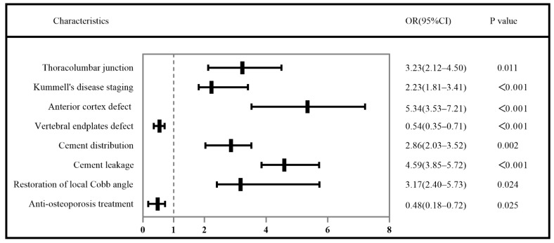
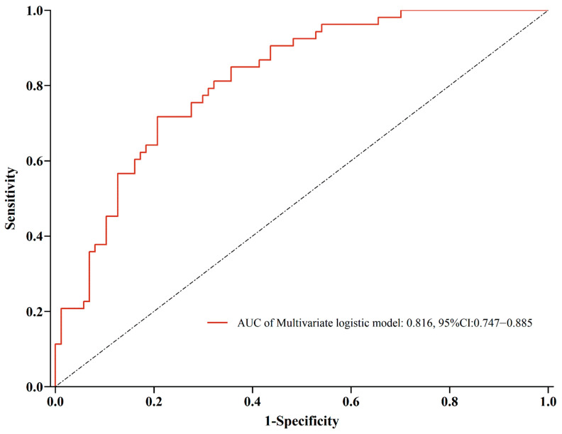
Objective: To investigate the independent influencing factors of bone cement displacement following percutaneous vertebral augmentation (PVA) in patients with stage I and stage II Kümmell’s disease. Methods: We retrospectively reviewed the records of 824 patients with stage Ⅰ and stage Ⅱ Kümmell’s disease treated with percutaneous vertebroplasty (PVP) or percutaneous vertebroplasty (PKP) from January 2016 to June 2022. Patients were divided into the postoperative bone cement displacement group (n = 150) and the bone cement non-displacement group (n = 674) according to the radiographic inspection results. The following data were collected: age, gender, body mass index (BMI), underlying disease, bone mineral density (BMD), involved vertebral segment, Kümmell’s disease staging, anterior height, local Cobb angle, the integrity of anterior vertebral cortex, the integrity of endplate in surgical vertebrae, surgical method, surgical approach, the volume of cement, distribution of cement, the viscosity of cement, cement leakage, and postoperative anti-osteoporosis treatment. Binary logistic regression analysis was performed to determine the independent influencing factors of bone cement displacement. The discrimination ability was evaluated using the area under the curve (AUC) of the receiver operating characteristic (ROC). Results: The results of logistic regression analysis revealed that thoracolumbar junction (odds ratio (OR) = 3.23, 95% confidence interval (CI) 2.12−4.50, p = 0.011), Kümmell’s disease staging (OR = 2.23, 95% CI 1.81−3.41, p < 0.001), anterior cortex defect (OR = 5.34, 95% CI 3.53−7.21, p < 0.001), vertebral endplates defect (OR = 0.54, 95% CI 0.35−0.71, p < 0.001), cement distribution (OR = 2.86, 95% CI 2.03−3.52, p = 0.002), cement leakage (OR = 4.59, 95% CI 3.85−5.72, p < 0.001), restoration of local Cobb angle (OR = 3.17, 95% CI 2.40−5.73, p = 0.024), and postoperative anti-osteoporosis treatment (OR = 0.48, 95% CI 0.18−0.72, p = 0.025) were independently associated with the bone cement displacement. The results of the ROC curve analysis showed that the AUC was 0.816 (95% CI 0.747−0.885), the sensitivity was 0.717, and the specificity was 0.793. Conclusion: Thoracolumbar fracture, stage Ⅱ Kümmell’s disease, anterior cortex defect, uneven cement distribution, cement leakage, and high restoration of the local Cobb angle were risk factors for cement displacement after PVA in Kümmell’s disease, while vertebral endplates defect and postoperative anti-osteoporosis treatment are protective factors.
Keywords: Kümmell’s disease; osteoporosis; percutaneous vertebral augmentation; postoperative complications; risk factors.
Conflict of interest statement
The authors declare no conflict of interest.
Figures
Similar articles
-
Comparative analysis of percutaneous vertebroplasty and kyphoplasty in the treatment of Stage III Kummell's disease without neurological symptoms: a retrospective study.J Orthop Surg Res. 2024 Aug 27;19(1):515. doi: 10.1186/s13018-024-05019-w. J Orthop Surg Res. 2024. PMID: 39192332 Free PMC article.
-
Risk factors for bone cement displacement after percutaneous vertebral augmentation for osteoporotic vertebral compression fractures.Front Surg. 2022 Jul 28;9:947212. doi: 10.3389/fsurg.2022.947212. eCollection 2022. Front Surg. 2022. PMID: 35965863 Free PMC article.
-
Analysis of the effect of percutaneous vertebroplasty in the treatment of thoracolumbar Kümmell's disease with or without bone cement leakage.BMC Musculoskelet Disord. 2021 Jan 5;22(1):10. doi: 10.1186/s12891-020-03901-2. BMC Musculoskelet Disord. 2021. PMID: 33402168 Free PMC article.
-
Consecutive Kummell's Disease Combined with Parkinson's Disease and Experienced Internal Fixation Failure: A Case Report and Literature Review.Orthop Surg. 2022 Jul;14(7):1533-1540. doi: 10.1111/os.13260. Epub 2022 May 27. Orthop Surg. 2022. PMID: 35633056 Free PMC article. Review.
-
Cluster phenomenon of vertebral refractures after posterior pedicle screw fixation in a patient with glucocorticosteroid-induced Kümmell's disease: a treatment dilemma.Arch Osteoporos. 2021 Jun 8;16(1):93. doi: 10.1007/s11657-021-00941-6. Arch Osteoporos. 2021. PMID: 34105042 Review.
Cited by
-
Comparative analysis of percutaneous vertebroplasty and kyphoplasty in the treatment of Stage III Kummell's disease without neurological symptoms: a retrospective study.J Orthop Surg Res. 2024 Aug 27;19(1):515. doi: 10.1186/s13018-024-05019-w. J Orthop Surg Res. 2024. PMID: 39192332 Free PMC article.
-
Clinical and radiographical analysis of percutaneous kyphoplasty with multi-point cement anchoring technique for preventing bone cement displacement in Kümmell's disease of stage I and II.Front Endocrinol (Lausanne). 2025 Jun 11;16:1538337. doi: 10.3389/fendo.2025.1538337. eCollection 2025. Front Endocrinol (Lausanne). 2025. PMID: 40568562 Free PMC article.
-
Ultrasonography-guided canal decompression combined with vertebroplasty and cement-augmented pedicle screw fixation for stage III Kümmell's disease with neurological deficits: a retrospective cohort study.BMC Musculoskelet Disord. 2024 Oct 12;25(1):805. doi: 10.1186/s12891-024-07929-6. BMC Musculoskelet Disord. 2024. PMID: 39395951 Free PMC article.
-
Percutaneous Transpedicular Intravertebral Cage Augmentation with Short-Segment Fixation Using Specially Designed Cannulated Cage Trials for Advanced Kümmell Disease: A Preliminary Study Comparing with Vertebroplasty with Short-Segment Fixation.Clin Orthop Surg. 2025 Feb;17(1):29-38. doi: 10.4055/cios24276. Epub 2025 Jan 14. Clin Orthop Surg. 2025. PMID: 39912073 Free PMC article.
-
Analysis of Risk Factors for Augmented Vertebral Refracture After Percutaneous Kyphoplasty in Osteoporotic Vertebral Compression Fractures.J Clin Med. 2025 Jan 8;14(2):329. doi: 10.3390/jcm14020329. J Clin Med. 2025. PMID: 39860335 Free PMC article.
References
-
- Adamska O., Modzelewski K., Stolarczyk A., Kseniuk J. Is Kummell’s Disease a Misdiagnosed and/or an Underreported Complication of Osteoporotic Vertebral Compression Fractures? A Pattern of the Condition and Available Treatment Modalities. J. Clin. Med. 2021;10:2584. doi: 10.3390/jcm10122584. - DOI - PMC - PubMed
Grants and funding
LinkOut - more resources
Full Text Sources
Miscellaneous
