

Primary Cutaneous B-Cell Lymphoma Co-Existing with Mycosis Fungoides-A Case Report and Overview of the Literature
- PMID: 36556432
- PMCID: PMC9785996
- DOI: 10.3390/life12122067
Primary Cutaneous B-Cell Lymphoma Co-Existing with Mycosis Fungoides-A Case Report and Overview of the Literature
Abstract
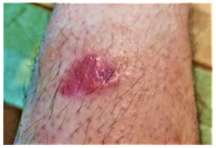
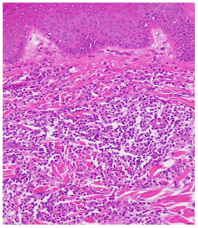
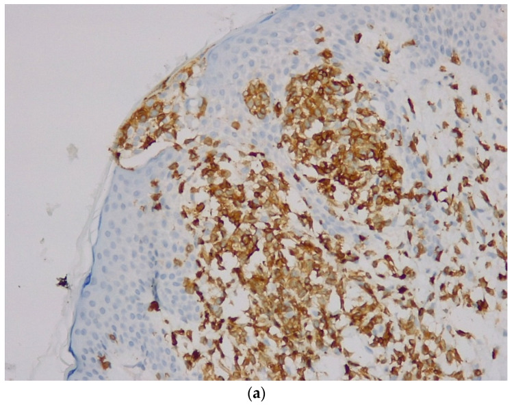
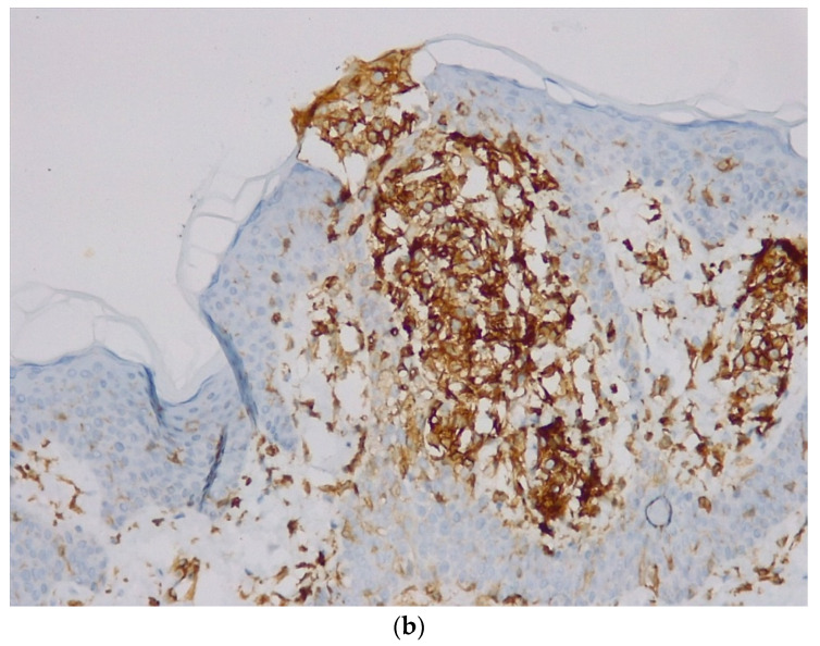
The existence of two sequential lymphomas, one localized and one systemic, either both with B or T lymphocytes, or one with B cells and one with T cells, with the same patient, is a known possibility. The second lymphoma is often induced by immunodepression or by the initial treatment. However, the existence of two cutaneous lymphomas with different cell lines, without systemic involvement, represents an uncommon situation. In this report, we describe the case of a 37-year-old man with an initial diagnosis of PMZBCL that over 10 months also developed a MF patch/plaque on the left leg.
Keywords: B-cell lymphoma; Mycosis Fungoides; composite lymphomas; primary cutaneous lymphoma.
Conflict of interest statement
The authors declare no conflict of interest.
Figures


Similar articles
-
Cutaneous lymphomas other than mycosis fungoides in Singapore: a clinicopathological analysis using recent classification systems.Br J Dermatol. 2003 Sep;149(3):542-53. doi: 10.1046/j.1365-2133.2003.05476.x. Br J Dermatol. 2003. PMID: 14510987
-
Mycosis fungoides: case report and literature review.Clin Med Insights Case Rep. 2014 Sep 3;7:95-8. doi: 10.4137/CCRep.S15724. eCollection 2014. Clin Med Insights Case Rep. 2014. PMID: 25232282 Free PMC article.
-
Full clinical recovery after topical acyclovir treatment of Epstein-Barr virus associated cutaneous B-cell lymphoma in patient with mycosis fungoides.Croat Med J. 2005 Jun;46(3):458-62. Croat Med J. 2005. PMID: 15861527
-
Primary cutaneous T-cell lymphoma (mycosis fungoides and Sézary syndrome): part I. Diagnosis: clinical and histopathologic features and new molecular and biologic markers.J Am Acad Dermatol. 2014 Feb;70(2):205.e1-16; quiz 221-2. doi: 10.1016/j.jaad.2013.07.049. J Am Acad Dermatol. 2014. PMID: 24438969 Review.
-
Non-mycosis fungoides cutaneous lymphomas in a referral center in Taiwan: A retrospective case series and literature review.PLoS One. 2020 Jan 24;15(1):e0228046. doi: 10.1371/journal.pone.0228046. eCollection 2020. PLoS One. 2020. PMID: 31978091 Free PMC article. Review.
Cited by
-
Immunosequencing applications in cutaneous T-cell lymphoma.Front Immunol. 2023 Dec 21;14:1300061. doi: 10.3389/fimmu.2023.1300061. eCollection 2023. Front Immunol. 2023. PMID: 38213330 Free PMC article. Review.
References
-
- Hoefnagel J.J., Vermeer M.H., Jansen P.M., Heule F., van Voorst Vader P.C., Sanders C.J., Gerritsen M.J., Geerts M.L., Meijer C.J., Noordijk E.M., et al. Primary Cutaneous Marginal Zone B-Cell Lymphoma: Clinical and Therapeutic Features in 50 Cases. Arch. Dermatol. 2005;141:1139–1145. doi: 10.1001/archderm.141.9.1139. - DOI - PubMed
Publication types
LinkOut - more resources
Full Text Sources
