Review
. 2022 Dec 12;12(12):2084.
doi: 10.3390/life12122084.

Thyroid Eye Disease

Affiliations
Review

Thyroid Eye Disease

Ramy Rashad et al. Life (Basel). .

Abstract

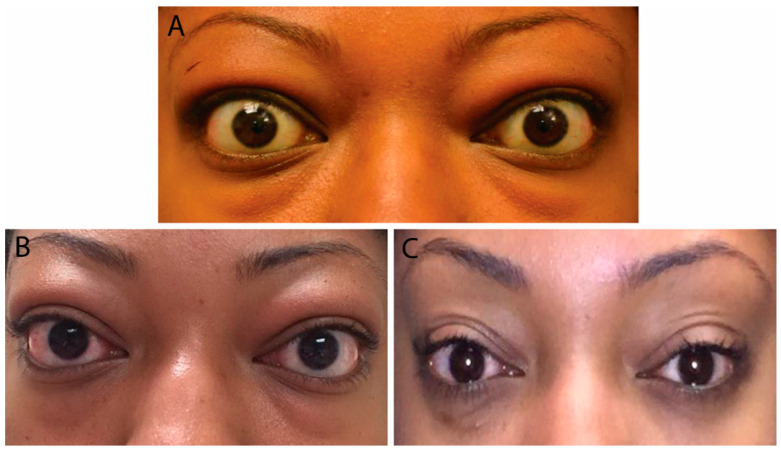
Thyroid eye disease (TED), an autoimmune inflammatory disorder of the orbit, presents with a potential array of clinical sequelae. The pathophysiology behind TED has been partially characterized in the literature. There remain certain elusive mechanisms welcoming of research advances. Disease presentation can vary, but those that follow a characteristic course start mild and increase in severity before plateauing into an inactive phase. Diagnosis and evaluation include careful physical examination, targeted laboratory work up, appropriate imaging studies, and tailored treatment regimens. Special consideration may apply to certain populations, such as pediatric and pregnant patients.

Keywords: Grave’s disease; Hashimoto’s thyroiditis; chronic lymphocytic thyroiditis; euthyroid eye disease; exophthalmos; orbital inflammation; proptosis; teprotumumab; thyroid eye disease; thyroid-associated orbitopathy.


Conflict of interest statement

The authors declare no conflict of interest.

Figures

Figure 1
Figure 1
Mild Thyroid Eye Disease. External color photograph showing mild signs of bilateral lower eyelid retraction, lateral upper eyelid flare, periorbital edema, and trace conjunctival injection.
Figure 2
Figure 2
External color photograph of a patient with chronic moderate thyroid eye disease with (A) upper and lower eyelid retraction, lateral flare of the upper eyelids, and moderate periorbital fat protrusion. (B) Proptosis, best seen in “worm’s eye view” (chin-up position).
Figure 3
Figure 3
(A) External color photograph of patient with acute moderate thyroid eye disease beginning two months after radioactive iodine therapy. (B) One month later with progression to severe thyroid eye disease. Exam shows increased conjunctival injection and chemosis. Sixty milligrams of oral prednisone with adjunctive radiotherapy was started. (C) Significant improvement in conjunctival and periorbital edema after one month. Steroids were slowly tapered over eight months.
Figure 4
Figure 4
(A) External color photograph of chronic moderate thyroid eye disease with asymmetric proptosis on left more than right. (B) Coronal CT of orbits with thickening of recti muscles on left more than right. (C) Sagittal cut of same CT scan.
Figure 5
Figure 5
(A) MRI orbits with gadolinium with sagittal cut showing thickening of bilateral medial recti muscles. (B) Coronal cut from same patient showing thickening of all bilateral inferior, medial, and superior recti muscles, as well as levator palpebrae superioris and superior oblique muscles.
Figure 6
Figure 6
(A) External color photograph of acute moderate thyroid eye disease with periorbital edema, upper and lower eyelid retraction, and chemosis (conjunctival swelling). (B) Same patient in chronic phase. (C) Patient status post-bilateral orbital fat decompression. No further procedures were needed.
Figure 7
Figure 7
External color photograph depicting a patient with strabismus. The right eye is slightly elevated and abducted in comparison to the left eye, as noted by the position of the corneal light reflex. A slight left head tilt is also taken to help combat the double vision. Right upper eyelid retraction is also noted.

Similar articles

Cited by

References

    1. Szczapa-Jagustyn J., Gotz-Wieckowska A., Kociecki J. An update on thyroid-associated ophthalmopathy in children and adolescents. J. Pediatr. Endocrinol. Metab. 2016;29:1115–1122. doi: 10.1515/jpem-2016-0122. - DOI - PubMed
    1. McAlinden C. An overview of thyroid eye disease. Eye Vis. 2014;1:9. doi: 10.1186/s40662-014-0009-8. - DOI - PMC - PubMed
    1. Yin X., Latif R., Bahn R., Davies T.F. Genetic profiling in Graves’ disease: Further evidence for lack of a distinct genetic contribution to Graves’ ophthalmopathy. Thyroid. 2012;22:730–736. doi: 10.1089/thy.2012.0007. - DOI - PMC - PubMed
    1. Stan M.N., Bahn R.S. Risk factors for development or deterioration of Graves’ ophthalmopathy. Thyroid. 2010;20:777–783. doi: 10.1089/thy.2010.1634. - DOI - PMC - PubMed
    1. Stan M.N. Natural History, Risk Factors, and Management of Patients with Mild GO. In: Bahn R.S., editor. Graves’ Disease—A Comprehensive Guide for Clinicians. Springer Science+Business Media; New York, NY, USA: 2015. pp. 241–255.

LinkOut - more resources