. 2022 Dec 14;12(12):2102.
doi: 10.3390/life12122102.

Effect of Photodynamic Therapy with Chlorin e6 on Canine Tumors

Affiliations

Effect of Photodynamic Therapy with Chlorin e6 on Canine Tumors

Rajeev Shrestha et al. Life (Basel). .

Abstract

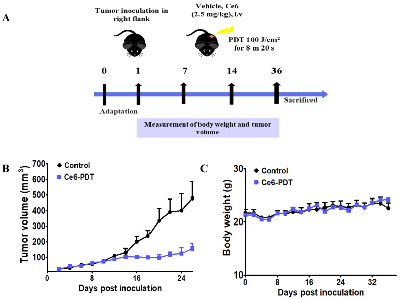
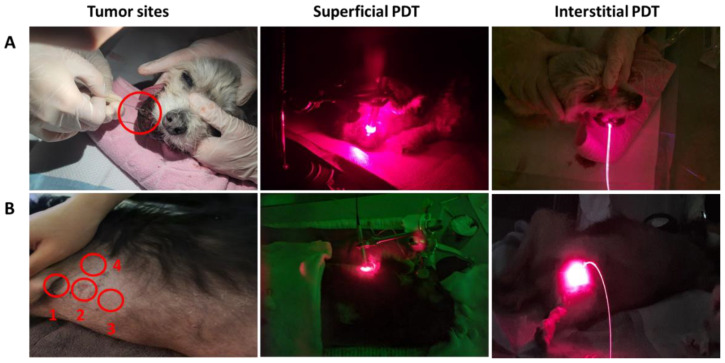
This work aims to prepare pure Chlorin e6 (Ce6) and establish Ce6-mediated photodynamic therapy (Ce6-PDT) as a better therapy option for canine tumors as well as mouse tumor models. Five dogs suffering from various cancers were treated with Ce6-PDT from one to several times. After receiving the Ce6 (2.5 mg/kg) for 3 h, tumors were illuminated superficially or interstitially with 660 nm light. Two dogs underwent Ce6-guided fluorescence imaging by photodynamic diagnosis (PDD). Cell proliferation and apoptosis were detected by the 4,5-dimethylthiazol-2-yl)-2,5-diphenyl tetrazolium bromide (MTT) assay and western blot assay, respectively. Ce6-PDT efficacy was also determined using melanoma and pancreatic cancer mouse models. Two veterinary patients with mammary carcinoma and histiocytic sarcoma had their tumors significantly diminished and showed improved health after receiving Ce6-PDT. Moreover, in the cases of canine tumors, the adjunctive use of Ce6-PDD revealed cancers that were not visible with white light viewing and provided a visual contrast from surrounding tissues. Also, in vivo, Ce6-PDT remarkably reduced melanoma and pancreatic tumors in the mouse model. These findings could pave the way for a better understanding of the underlying processes of Ce6-PDT, making it an effective and safe candidate for use in human and veterinary applications to abolish various cancers.

Keywords: Ce6; PDD; PDT; canine tumors; photosensitizer.


Conflict of interest statement

The authors declare no conflict of interest.

Figures

Scheme 1
Scheme 1
Synthesis of Ce6 and its PDT or PDD effects on veterinary cases, in vitro, and in vivo.
Figure 1
Figure 1
Absorbance and fluorescence of Ce6 in 95% EtOH solution (10 µM).
Figure 2
Figure 2
Ce6-mediated cytotoxicity on human pancreatic cancer cells. (A) Fluorescence images of MIA PaCa-2 cells after 24 h incubation with Ce6. The images shown are the white light images (left), 400 nm (middle), and merged of two images (right). The scale bar represents 100 μm. (B,C) Dark and photo-induced cytotoxic activity of Ce6 against AsPC-1 and MIA PaCa-2 cell lines (equivalent concentration 0–512 μΜ for dark and 0–51.2 μΜ of Ce6 for light). Medium with laser was taken as a control group. The data are the representation of mean ± SEM obtained from three independent experiments. (D,E) Morphological changes in cells after Ce6 treatment.
Figure 3
Figure 3
Ce6-mediated PDT effects in protein levels of apoptosis. AsPC-1 and MIA PaCa-2 cells were treated with Ce6 (5 and 10 μM) for 3 h followed by or without PDT. The western blot assay detected the protein levels of Bcl-2, Bax, Procaspase-3, cleaved caspase-3, and cleaved PARP-1 in (A). AsPC-1 and (B) MIA PaCa-2 cell line. β-Actin was used as a loading control.
Figure 4
Figure 4
Anti-cancer activities of Ce6-PDT against melanoma allografts. (A) Timeline of the experimental procedure. The mice were subcutaneously injected at the right flanks with 0.1 mL of B16F10 cells (1 × 10⁷ cells/mL). The control group was not exposed to any treatment. (B) Changes in tumor volume and (C) body weight for control and treatment groups. Data are expressed as mean ± SEM, n = 5.
Figure 4
Figure 4
Anti-cancer activities of Ce6-PDT against melanoma allografts. (A) Timeline of the experimental procedure. The mice were subcutaneously injected at the right flanks with 0.1 mL of B16F10 cells (1 × 10⁷ cells/mL). The control group was not exposed to any treatment. (B) Changes in tumor volume and (C) body weight for control and treatment groups. Data are expressed as mean ± SEM, n = 5.
Figure 5
Figure 5
In vivo therapeutic effect in PANC02 tumor models. (A) Experimental scheme of tumor induction and Ce6-PDT treatment. (B) Tumor volume variations of each group over time. (C) Body weight variations of each group over time. Data are expressed as mean ± SEM, n = 5.
Figure 6
Figure 6
Photodynamic diagnosis (PDD) cases with representative images. The dog was intravenously injected with 2.5 mg/kg Ce6. After 3 h, prior to the PDT, fluorescence was detected in dog’s abdominal tumor and normal groin skin (A) with white light and (B) blue light at 405 nm. In the blue light, intense fluorescence was detected at tumor locations, whereas normal tissues had no fluorescence.
Figure 7
Figure 7
Photodynamic therapy of case no. 4 and 5. (A) PDT on case 4 at oral mucosa through the superficial and interstitial method. (B) PDT on case 5 at the abdominal and groin skin through the superficial and interstitial methods. The patients were intravenously injected with 2.5 mg/kg Ce6. After 3 h, the tumor was firstly treated with superficial irradiation by using the quartz fiber fitted with a macrolens which was set toward the surface of the tumor. For the interstitial irradiation, an over-the-needle intravenous catheter (19 gauge needle) was inserted at the desired location of the tumor mass. In both methods, the light irradiation was 660 nm and emitted (200 mW, 100 J) by a laser diode under general anesthesia. The treatment was processed for 500 s in repeated manners.
Figure 8
Figure 8
Computed tomography (CT) images of dog 5. Dog 5 was diagnosed with histiocytic sarcoma at left axillary skin, abdominal and groin skin before first PDT. (A) Opacities of tumors areas could be observed in the images of the axillary skin (red arrow), (B) After second PDT treatment, the tumor on axillary skin was significantly diminished in size and the margin of the mass became unclear.

Similar articles

Cited by

References

    1. Gunaydin G., Gedik M.E., Ayan S. Photodynamic Therapy for the Treatment and Diagnosis of Cancer–A Review of the Current Clinical Status. Front. Chem. 2021;9:686303. doi: 10.3389/fchem.2021.686303. - DOI - PMC - PubMed
    1. Simelane N.W.N., Kruger C.A., Abrahamse H. Photodynamic diagnosis and photodynamic therapy of colorectal cancer in vitro and in vivo. RSC Adv. 2020;10:41560–41576. doi: 10.1039/D0RA08617G. - DOI - PMC - PubMed
    1. Xu S., Bulin A.L., Hurbin A., Elleaume H., Coll J.L., Broekgaarden M. Photodynamic Diagnosis and Therapy for Peritoneal Carcinomatosis: Emerging Perspectives. Cancers. 2020;12:2491. doi: 10.3390/cancers12092491. - DOI - PMC - PubMed
    1. Miyake M., Nishimura N., Nakai Y., Fujii T., Owari T., Hori S., Morizawa Y., Gotoh D., Anai S., Torimoto K., et al. Photodynamic diagnosis-assisted transurethral resection using oral 5-aminolevulinic acid decreases the risk of repeated recurrence in non-muscle-invasive bladder cancer: A cumulative incidence analysis by the person-time method. Diagnostics. 2021;11:185. doi: 10.3390/diagnostics11020185. - DOI - PMC - PubMed
    1. Castano A.P., Demidova T.N., Hamblin M.R. Mechanisms in photodynamic therapy: Part one-photosensitizers, photochemistry and cellular localization. Photodiagn. Photodyn. Ther. 2004;1:279–293. doi: 10.1016/S1572-1000(05)00007-4. - DOI - PMC - PubMed

LinkOut - more resources