

Osteogenic Potential of Monosodium Urate Crystals in Synovial Mesenchymal Stem Cells
- PMID: 36556927
- PMCID: PMC9786019
- DOI: 10.3390/medicina58121724
Osteogenic Potential of Monosodium Urate Crystals in Synovial Mesenchymal Stem Cells
Abstract
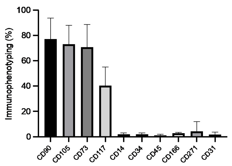
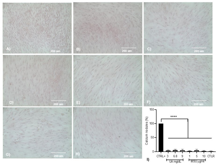
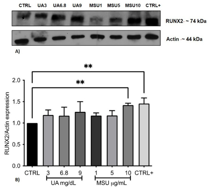
Background and Objectives: Deposits of monosodium urate (MSU) crystals due to increased levels of uric acid (UA) have been associated with bone formation and erosion, mainly in patients with chronic gout. The synovial membrane (SM) comprises several types of cells, including mesenchymal stem cells (SM-MSCs); however, it is unknown whether UA and MSU induce osteogenesis through SM-MSCs. Materials and Methods: Cultures of SM were immunotyped with CD44, CD69, CD90, CD166, CD105, CD34, and CD45 to identify MSCs. CD90+ cells were isolated by immunomagnetic separation (MACS), colony-forming units (CFU) were identified, and the cells were exposed to UA (3, 6.8, and 9 mg/dL) and MSU crystals (1, 5, and 10 μg/mL) for 3 weeks, and cellular morphological changes were evaluated. IL-1β and IL-6 were determined by ELISA, mineralization was assessed by alizarin red, and the expression of Runx2 was assessed by Western blot. Results: Cells derived from SM and after immunomagnetic separation were positive for CD90 (53 ± 8%) and CD105 (52 ± 18%) antigens, with 53 ± 5 CFU identified. Long-term exposure to SM-MSCs by UA and MSU crystals did not cause morphological damage or affect cell viability, nor were indicators of inflammation detected. Mineralization was observed at doses of 6.8 mg/dL UA and 5 μg/mL MSU crystals; however, the differences were not significant with respect to the control. The highest dose of MSU crystals (10 μg/mL) induced significant Runx2 expression with respect to the control (1.4 times greater) and SM-MSCs cultured in the osteogenic medium. Conclusions: MSU crystals may modulate osteogenic differentiation of SM-MSCs through an increase in Runx2.
Keywords: gout; mesenchymal stem cells; monosodium urate crystals; osteodifferentiation; synovial membrane.
Conflict of interest statement
The authors declare no conflict of interest with respect to the research, authorship, and/or publication of this study.
Figures


Similar articles
-
Factors secreted by monosodium urate crystal-stimulated macrophages promote a proinflammatory state in osteoblasts: a potential indirect mechanism of bone erosion in gout.Arthritis Res Ther. 2022 Sep 5;24(1):212. doi: 10.1186/s13075-022-02900-z. Arthritis Res Ther. 2022. PMID: 36064735 Free PMC article.
-
Monosodium urate monohydrate crystals inhibit osteoblast viability and function: implications for development of bone erosion in gout.Ann Rheum Dis. 2011 Sep;70(9):1684-91. doi: 10.1136/ard.2010.144774. Epub 2011 May 27. Ann Rheum Dis. 2011. PMID: 21622970
-
Macrophage-derived IL-1β enhances monosodium urate crystal-triggered NET formation.Inflamm Res. 2017 Mar;66(3):227-237. doi: 10.1007/s00011-016-1008-0. Epub 2016 Nov 16. Inflamm Res. 2017. PMID: 27853847 Free PMC article.
-
The crystallization of monosodium urate.Curr Rheumatol Rep. 2014 Feb;16(2):400. doi: 10.1007/s11926-013-0400-9. Curr Rheumatol Rep. 2014. PMID: 24357445 Free PMC article. Review.
-
Urate Crystals; Beyond Joints.Front Med (Lausanne). 2021 Jun 4;8:649505. doi: 10.3389/fmed.2021.649505. eCollection 2021. Front Med (Lausanne). 2021. PMID: 34150794 Free PMC article. Review.
Cited by
-
Osteogenic Potential and Bone Matrix Maturity: Comparison of Demineralized Bone Matrix and P15 Polypeptide iFactor® in an In Vitro Study.Medicina (Kaunas). 2025 May 18;61(5):914. doi: 10.3390/medicina61050914. Medicina (Kaunas). 2025. PMID: 40428872 Free PMC article.
References
-
- Shi D., Chen J.Y., Wu H.X., Zhou Q.J., Chen H.Y., Lu Y.F., Yu R.S. Relationship between urate within tophus and bone erosion according to the anatomic location of urate deposition in gout: A quantitative analysis using dual-energy CT volume measurements. Medicine. 2019;98:e18431. doi: 10.1097/MD.0000000000018431. - DOI - PMC - PubMed
-
- McQueen F.M., Doyle A., Reeves Q., Gao A., Tsai A., Gamble G.D., Curteis B., Williams M., Dalbeth N. Bone erosions in patients with chronic gouty arthropathy are associated with tophi but not bone oedema or synovitis: New insights from a 3 T MRI study. Rheumatol. 2014;53:95–103. doi: 10.1093/rheumatology/ket329. - DOI - PubMed
-
- Dalbeth N., Smith T., Nicolson B., Clark B., Callon K., Naot D., Haskard D.O., McQueen F.M., Reid I.R., Cornish J. Enhanced osteoclastogenesis in patients with tophaceous gout: Urate crystals promote osteoclast development through interactions with stromal cells. Arthritis. Rheum. 2008;58:1854–1865. doi: 10.1002/art.23488. - DOI - PubMed
MeSH terms
Substances
LinkOut - more resources
Full Text Sources
Medical
Research Materials
Miscellaneous
