

Vascular Diameter as Clue for the Diagnosis of Clinically and/or Dermoscopically Equivocal Pigmented and Non-Pigmented Basal Cell Carcinomas and Nodular Melanomas
- PMID: 36556965
- PMCID: PMC9786710
- DOI: 10.3390/medicina58121761
Vascular Diameter as Clue for the Diagnosis of Clinically and/or Dermoscopically Equivocal Pigmented and Non-Pigmented Basal Cell Carcinomas and Nodular Melanomas
Abstract
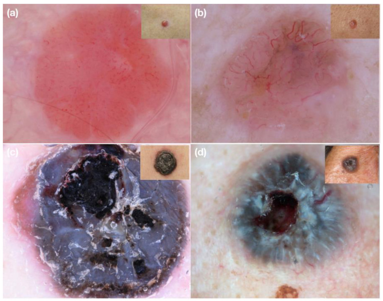
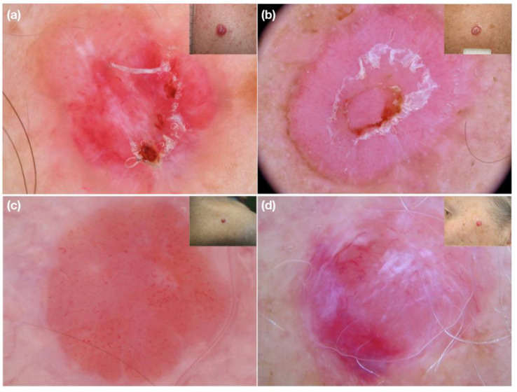
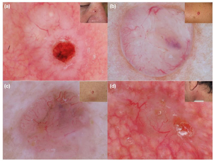
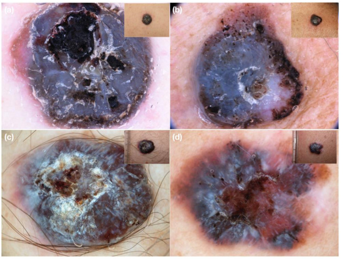
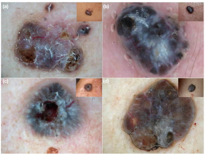
Background and objectives: Dermoscopy is a useful tool for the early and non-invasive diagnosis of skin malignancies. Besides many progresses, heavily pigmented and amelanotic skin tumors remain still a challenge. We aimed to investigate by dermoscopy if distinctive morphologic characteristics of vessels may help the diagnosis of equivocal nodular lesions. Materials and Methods: A collage of 16 challenging clinical and dermoscopic images of 8 amelanotic and 8 heavily pigmented nodular melanomas and basal cell carcinomas was sent via e-mail to 8 expert dermoscopists. Results: Dermoscopy improved diagnostic accuracy in 40 cases. Vessels were considered the best clue in 71 cases. Focusing on the diameter of vessels improved diagnosis in 5 cases. Conclusions: vascular diameter in addition to morphology and arrangement may be a useful dermoscopic clue for the differential diagnosis of clinically equivocal nodular malignant tumors.
Keywords: basal cell carcinoma; dermoscopy; melanoma; skin cancer; vascular diameter; vessels.
Conflict of interest statement
The authors declare no conflict of interest.
Figures
Similar articles
-
Dermoscopy improves diagnostic accuracy for clinically amelanotic nodules.Australas J Dermatol. 2019 Feb;60(1):45-49. doi: 10.1111/ajd.12902. Epub 2018 Aug 19. Australas J Dermatol. 2019. PMID: 30123971
-
Accuracy of Dermoscopic Criteria for the Diagnosis of Melanoma In Situ.JAMA Dermatol. 2018 Apr 1;154(4):414-419. doi: 10.1001/jamadermatol.2017.6447. JAMA Dermatol. 2018. PMID: 29466542 Free PMC article.
-
Updates in Dermoscopy for Pigmented Lesions.Dermatol Clin. 2025 Jul;43(3):419-432. doi: 10.1016/j.det.2025.03.009. Epub 2025 May 12. Dermatol Clin. 2025. PMID: 40581422 Review.
-
Pigmented nodular melanoma: the predictive value of dermoscopic features using multivariate analysis.Br J Dermatol. 2015 Jul;173(1):106-14. doi: 10.1111/bjd.13861. Epub 2015 Jun 2. Br J Dermatol. 2015. PMID: 25916655
-
Clinical and dermoscopic characteristics of amelanotic melanomas that are not of the nodular subtype.J Eur Acad Dermatol Venereol. 2012 May;26(5):591-6. doi: 10.1111/j.1468-3083.2011.04122.x. Epub 2011 May 18. J Eur Acad Dermatol Venereol. 2012. PMID: 21585561 Review.
Cited by
-
Dermoscopy of Basal Cell Carcinoma Part 3: Differential Diagnosis, Treatment Monitoring and Novel Technologies.Cancers (Basel). 2025 Mar 19;17(6):1025. doi: 10.3390/cancers17061025. Cancers (Basel). 2025. PMID: 40149358 Free PMC article. Review.
References
-
- Argenziano G., Longo C., Cameron A., Cavicchini S., Gourhant J.Y., Lallas A., McColl I., Rosendahl C., Thomas L., Tiodorovic-Zivkovic D., et al. Blue-black rule: A simple dermoscopic clue to recognize pigmented nodular melanoma. Br. J. Dermatol. 2011;165:1251–1255. doi: 10.1111/j.1365-2133.2011.10621.x. - DOI - PubMed
MeSH terms
LinkOut - more resources
Full Text Sources
Medical
