

Platelet-Rich Plasma for Pleurodesis: An Experimental Study in Rabbits
- PMID: 36557044
- PMCID: PMC9785005
- DOI: 10.3390/medicina58121842
Platelet-Rich Plasma for Pleurodesis: An Experimental Study in Rabbits
Abstract
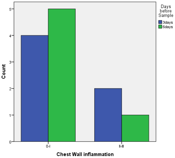
Background and Objectives: This study was designed to evaluate platelet-rich plasma (PRP) as a method of pleurodesis in a rabbit model. Pleurodesis with PRP was compared against the gold-standard use of talc. The secondary evaluation assessed the ideal time for achieving pleurodesis. Materials and Methods: 25 healthy New Zealand white rabbits were assigned to three groups, as follows: 12 animals in the first and second groups, as well as one animal with no intervention in the final group, which was used as a control. The talc pleurodesis group (baseline) underwent pleurodesis with sterile talc, which is the gold-standard sclerosing agent used for pleurodesis. The PRP group underwent pleurodesis using autologous PRP. The last group had one rabbit with no intervention. A total of 12 rabbits (n = 6 for the talc pleurodesis group and n = 6 for the PRP group) were sacrificed 3 days (72 h) after the intervention, and 12 rabbits (n = 6 for the talc pleurodesis group and n = 6 for the PRP group) were sacrificed 6 days (144 h) after the intervention. In both the talc and PRP group, FBC and CRP were measured before the intervention and in 3 or 6 days afterwards, respectively. The pleura and the lungs were evaluated histopathologically. Results: Macroscopically, there were no statistically significant differences between the two groups. In terms of microscopic findings, there were no statistically significant differences in inflammatory reactions provoked in the visceral and parietal pleura between the PRP and talc. In addition, with talc pleurodesis, a foreign-body reaction was observed in about 50% of the cases, which was not observed with PRP. In terms of inflammation between 3 and 6 days, there were no statistically significant differences with PRP, there was only a statistically significant difference between 3 and 6 days regarding the parietal pleura in the talc group. Conclusions: The instillation of autologous PRP in the pleural cavity shows promise in achieving pleurodesis. The efficacy of PRP as a pleurodesis agent should be examined further.
Keywords: PRP; pleurodesis; rabbits; talc.
Conflict of interest statement
The authors declare no conflict of interest.
Figures


Similar articles
-
A comparison of the effectiveness of talc, polidocanol and ethanol as pleural sclerosing agents in rabbits.Thorac Cardiovasc Surg. 2009 Mar;57(2):102-6. doi: 10.1055/s-2008-1039103. Epub 2009 Feb 24. Thorac Cardiovasc Surg. 2009. PMID: 19241312
-
Comparisons of pleurodesis induced by talc with or without thymol iodide in rabbits.Chest. 1998 Mar;113(3):795-9. doi: 10.1378/chest.113.3.795. Chest. 1998. PMID: 9515859
-
Malignant pleural effusion and talc pleurodesis. Experimental model regarding early kinetics of talc particle dissemination in the chest after experimental talc pleurodesis.J BUON. 2009 Jul-Sep;14(3):419-23. J BUON. 2009. PMID: 19810132
-
Mechanisms of pleurodesis.Respiration. 2012;83(2):91-8. doi: 10.1159/000335419. Epub 2012 Jan 20. Respiration. 2012. PMID: 22286268 Review.
-
Inflammation and clinical repercussions of pleurodesis induced by intrapleural talc administration.Clinics (Sao Paulo). 2007 Oct;62(5):627-34. doi: 10.1590/s1807-59322007000500015. Clinics (Sao Paulo). 2007. PMID: 17952325 Review.
References
MeSH terms
Substances
LinkOut - more resources
Full Text Sources
Research Materials
Miscellaneous
