. 2022 Dec 17;12(12):1278.
doi: 10.3390/membranes12121278.

Structural Role of Plasma Membrane Sterols in Osmotic Stress Tolerance of Yeast Saccharomyces cerevisiae

Affiliations

Structural Role of Plasma Membrane Sterols in Osmotic Stress Tolerance of Yeast Saccharomyces cerevisiae

Svyatoslav S Sokolov et al. Membranes (Basel). .

Abstract

Yeast S. cerevisiae has been shown to suppress a sterol biosynthesis as a response to hyperosmotic stress. In the case of sodium stress, the failure to suppress biosynthesis leads to an increase in cytosolic sodium. The major yeast sterol, ergosterol, is known to regulate functioning of plasma membrane proteins. Therefore, it has been suggested that the suppression of its biosynthesis is needed to adjust the activity of the plasma membrane sodium pumps and channels. However, as the sterol concentration is in the range of thirty to forty percent of total plasma membrane lipids, it is believed that its primary biological role is not regulatory but structural. Here we studied how lowering the sterol content affects the response of a lipid bilayer to an osmotic stress. In accordance with previous observations, we found that a decrease of the sterol fraction increases a water permeability of the liposomal membranes. Yet, we also found that sterol-free giant unilamellar vesicles reduced their volume during transient application of the hyperosmotic stress to a greater extent than the sterol-rich ones. Furthermore, our data suggest that lowering the sterol content in yeast cells allows the shrinkage to prevent the osmotic pressure-induced plasma membrane rupture. We also found that mutant yeast cells with the elevated level of sterol accumulated propidium iodide when exposed to mild hyperosmotic conditions followed by hypoosmotic stress. It is likely that the decrease in a plasma membrane sterol content stimulates a drop in cell volume under hyperosmotic stress, which is beneficial in the case of a subsequent hypo-osmotic one.

Keywords: giant unilamellar vesicle; hyperosmotic stress; hypoosmotic stress; large unilamellar vesicle; light scattering; sterol; yeast.


Conflict of interest statement

The authors declare no conflict of interest. The funders had no role in the design of the study; in the collection, analyses, or interpretation of data; in the writing of the manuscript; or in the decision to publish the results.

Figures

Figure 1
Figure 1
A scheme illustrating how sterol might provoke the pore formation upon changes in the osmotic pressure. High rigidity and low water permeability provided by high sterol might prevent the volume decrease upon high salt stress. In the case the hyperosmotic stress is followed by the hypoosmotic one, sterol-rich structures appear to be prone to rupturing. See text (Introduction) for details.
Figure 2
Figure 2
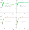
Representative dependences I(t) of the light intensity scattered from LUV suspensions after application of the hyperosmotic gradient. (a,b)—LUVs made from PLE (no cholesterol) with sorbitol (a) or sucrose (b) used as an osmolyte. (c,d)—LUVs made from PLE + cholesterol lipid mixture with sorbitol (c) or sucrose (d) used as an osmolyte. Solid blue and red curves represent the fit of the experimentally determined dependence I(t) (green traces) by Equation (3). The membrane permeability with respect to water determined from the fits is Pf = 5.7 µm/s (PLE + cholesterol membrane) and Pf = 12.7 µm/s (PLE membrane) both for sucrose and sorbitol used as an osmolyte. All curves are rather well fitted by a single-component Lambert function.
Figure 3
Figure 3
Time course of the GUV reaction on the transient hyperosmotic stress. (A)—GUV formed from DOPC; (B)—GUV formed from 70 mol.% DOPC + 30 mol.% Ergosterol; (C)—GUV formed from 70 mol.% DOPC + 30 mol.% Cholesterol. Time in seconds passed from the application of the hyperosmotic stress is indicated in left upper corners. Time count starts from the moment of the application of the hyperosmotic solution. Scale bar is 10 μm.
Figure 4
Figure 4
S. cerevisiae mutants with elevated PM sterol (Δerg4Δlam2, upc2-1 and upc2-1Δlam2) are more sensitive to hyperosmotic stress induced by 0.6 M NaCl, but not 0.6 M KCl or 1.2 M sorbitol. Y-axis: growth rate, division time equals 0.434/µ. * p < 0.05, Two-sample t-test for independent samples.
Figure 5
Figure 5
S. cerevisiaeΔlam1Δlam2Δlam3Δlam4 (lam 1234) mutant cells accumulate propidium iodide after hypoosmotic stress. Circles are individual experimental repeat measurements. * p < 0.05, Two-sample t-test for independent samples.

References

    1. Montañés F.M., Pascual-Ahuir A., Proft M. Repression of ergosterol biosynthesis is essential for stress resistance and is mediated by the Hog1 MAP kinase and the Mot3 and Rox1 transcription factors. Mol. Microbiol. 2011;79:1008–1023. doi: 10.1111/j.1365-2958.2010.07502.x. - DOI - PubMed
    1. Jordá T., Puig S. Regulation of ergosterol biosynthesis in Saccharomyces cerevisiae. Genes. 2020;11:795. doi: 10.3390/genes11070795. - DOI - PMC - PubMed
    1. Kodedová M., Sychrová H. Changes in the sterol composition of the plasma membrane affect membrane potential, salt tolerance and the activity of multidrug resistance pumps in Saccharomyces cerevisiae. PLoS ONE. 2015;10:e0139306. doi: 10.1371/journal.pone.0139306. - DOI - PMC - PubMed
    1. Dupont S., Beney L., Ferreira T., Gervais P. Nature of sterols affects plasma membrane behavior and yeast survival during dehydration. Biochim. Biophys. Acta. 2011;1808:1520–1528. doi: 10.1016/j.bbamem.2010.11.012. - DOI - PubMed
    1. Boudreault F., Tan J.J., Grygorczyk R. Propidium uptake and ATP release in A549 cells share similar transport mechanisms. Biophys. J. 2022;121:1593–1609. doi: 10.1016/j.bpj.2022.04.007. - DOI - PMC - PubMed

LinkOut - more resources