

An NMR-Based Model to Investigate the Metabolic Phenoreversion of COVID-19 Patients throughout a Longitudinal Study
- PMID: 36557244
- PMCID: PMC9788519
- DOI: 10.3390/metabo12121206
An NMR-Based Model to Investigate the Metabolic Phenoreversion of COVID-19 Patients throughout a Longitudinal Study
Abstract
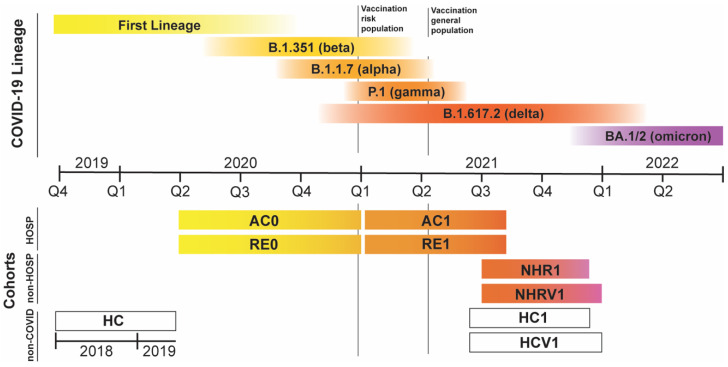
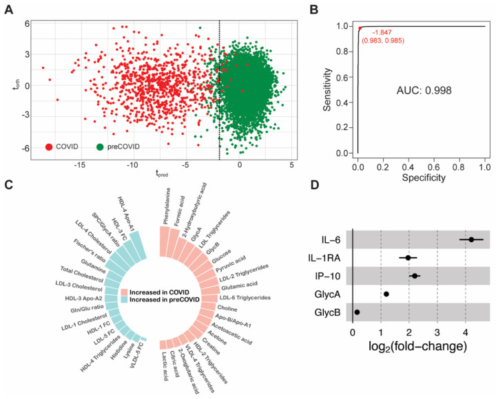
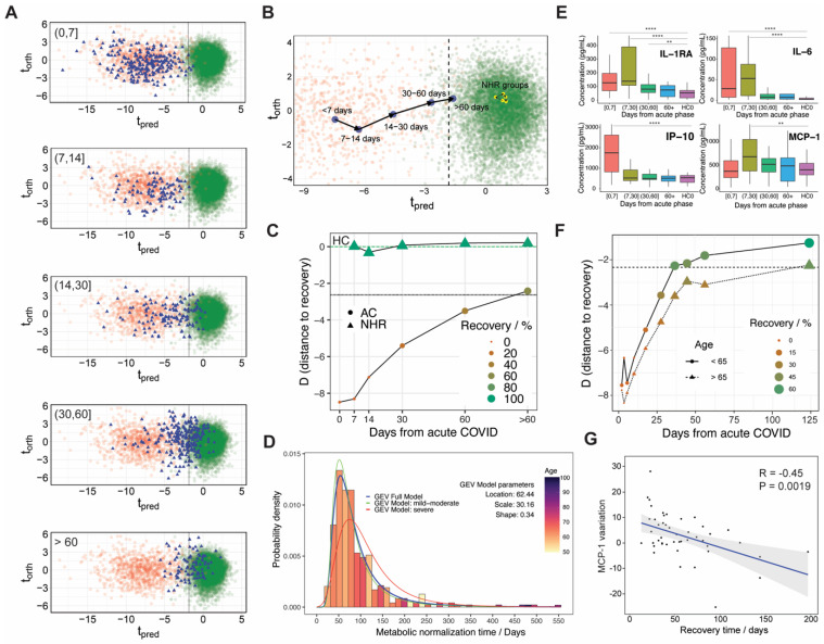
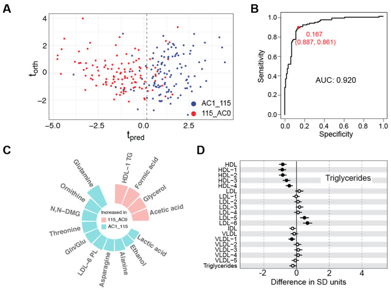
After SARS-CoV-2 infection, the molecular phenoreversion of the immunological response and its associated metabolic dysregulation are required for a full recovery of the patient. This process is patient-dependent due to the manifold possibilities induced by virus severity, its phylogenic evolution and the vaccination status of the population. We have here investigated the natural history of COVID-19 disease at the molecular level, characterizing the metabolic and immunological phenoreversion over time in large cohorts of hospitalized severe patients (n = 886) and non-hospitalized recovered patients that self-reported having passed the disease (n = 513). Non-hospitalized recovered patients do not show any metabolic fingerprint associated with the disease or immune alterations. Acute patients are characterized by the metabolic and lipidomic dysregulation that accompanies the exacerbated immunological response, resulting in a slow recovery time with a maximum probability of around 62 days. As a manifestation of the heterogeneity in the metabolic phenoreversion, age and severity become factors that modulate their normalization time which, in turn, correlates with changes in the atherogenesis-associated chemokine MCP-1. Our results are consistent with a model where the slow metabolic normalization in acute patients results in enhanced atherosclerotic risk, in line with the recent observation of an elevated number of cardiovascular episodes found in post-COVID-19 cohorts.
Keywords: COVID-19; atherosclerotic risk; inflammation; lipidomics; long COVID; metabolomics.
Conflict of interest statement
The authors declare no conflict of interest.
Figures
References
-
- Marín-Corral J., Rodríguez-Morató J., Gomez-Gomez A., Pascual-Guardia S., Muñoz-Bermúdez R., Salazar-Degracia A., Pérez-Terán P., Restrepo M.I., Khymenets O., Haro N., et al. Metabolic Signatures Associated with Severity in Hospitalized COVID-19 Patients. Int. J. Mol. Sci. 2021;22:4794. doi: 10.3390/ijms22094794. - DOI - PMC - PubMed
-
- Bruzzone C., Bizkarguenaga M., Gil-Redondo R., Diercks T., Arana E., García de Vicuña A., Seco M., Bosch A., Palazón A., San Juan I., et al. SARS-CoV-2 Infection Dysregulates the Metabolomic and Lipidomic Profiles of Serum. iScience. 2020;23:101645. doi: 10.1016/j.isci.2020.101645. - DOI - PMC - PubMed
-
- Fraser D.D., Slessarev M., Martin C.M., Daley M., Patel M.A., Miller M.R., Patterson E.K., O’Gorman D.B., Gill S.E., Wishart D.S., et al. Metabolomics Profiling of Critically Ill Coronavirus Disease 2019 Patients: Identification of Diagnostic and Prognostic Biomarkers. Crit. Care Explor. 2020;2:e0272. doi: 10.1097/CCE.0000000000000272. - DOI - PMC - PubMed
-
- Schmelter F., Föh B., Mallagaray A., Rahmöller J., Ehlers M., Lehrian S., von Kopylow V., Künsting I., Lixenfeld A.S., Martin E., et al. Metabolic and Lipidomic Markers Differentiate COVID-19 from Non-Hospitalized and Other Intensive Care Patients. Front. Mol. Biosci. 2021;8:1091. doi: 10.3389/fmolb.2021.737039. - DOI - PMC - PubMed
-
- Kimhofer T., Lodge S., Whiley L., Gray N., Loo R.L., Lawler N.G., Nitschke P., Bong S.-H., Morrison D.L., Begum S., et al. Integrative Modeling of Quantitative Plasma Lipoprotein, Metabolic, and Amino Acid Data Reveals a Multiorgan Pathological Signature of SARS-CoV-2 Infection. J. Proteome Res. 2020;19:4442–4454. doi: 10.1021/acs.jproteome.0c00519. - DOI - PubMed
Grants and funding
- the SPRI I + D COVID-19 fund (Basque Government, bG-COVID-19)
- BIOEF EITB Maratoia (BIO21/COV/037)
- the European Research Council (ERC) (ERC-2018-StG 804236-NEXTGEN-IO), ISCiii DTS21/00094, RYC2018-024183-I; and PID2019-107956RA-I00
- The Spinnaker Health Research Foundation, WA, The McCusker Foundation, WA, The Western Australian State Government
- MRFF (grant number 2014349) for funding the Australian National Phenome Centre
- the UK MRC
- the Department of Jobs, Tourism, Science and Innovation, Government of Western Australian Premier's Fellowship
- ARC Laureate Fellowship
- Western Australian Covid Research Response team
- Ministerio de Ciencia, Tecnología e Innovación (Minciencias), Ministerio de Educación Nacional, Ministerio de Industria, Comercio y Turismo e ICETEX (792-2017) 2a Convocatoria Ecosistema Científico - Colombia Científica para la Financiación de Proye
- World Bank and Vicerrectoría de Investigaciones, Pontificia Universidad Javeriana, Bogotá, Co-lombia (contract no. FP44842 - 221-2018)
LinkOut - more resources
Full Text Sources
Research Materials
Miscellaneous
