

Red Sea Atlas of Coral-Associated Bacteria Highlights Common Microbiome Members and Their Distribution across Environmental Gradients-A Systematic Review
- PMID: 36557593
- PMCID: PMC9787610
- DOI: 10.3390/microorganisms10122340
Red Sea Atlas of Coral-Associated Bacteria Highlights Common Microbiome Members and Their Distribution across Environmental Gradients-A Systematic Review
Abstract
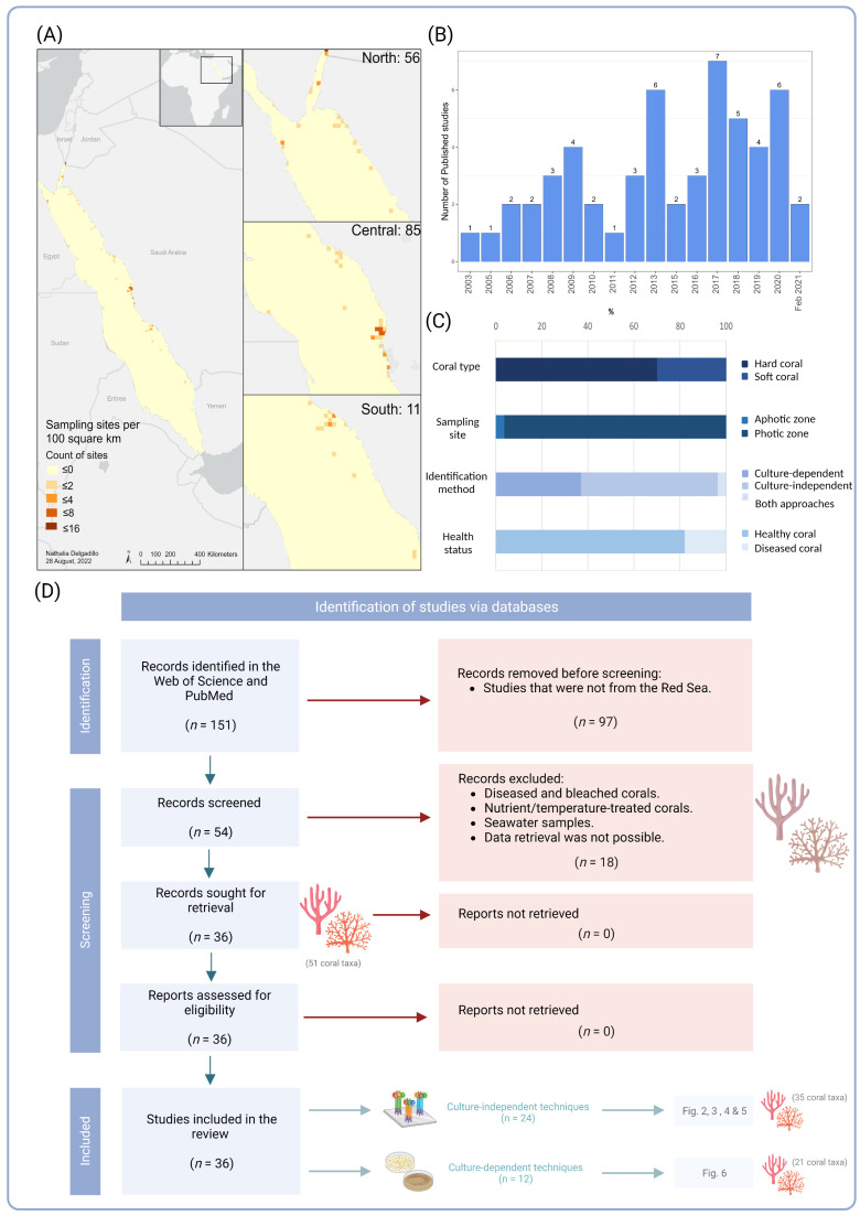
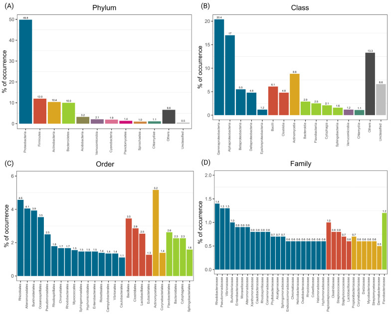
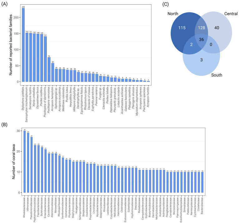
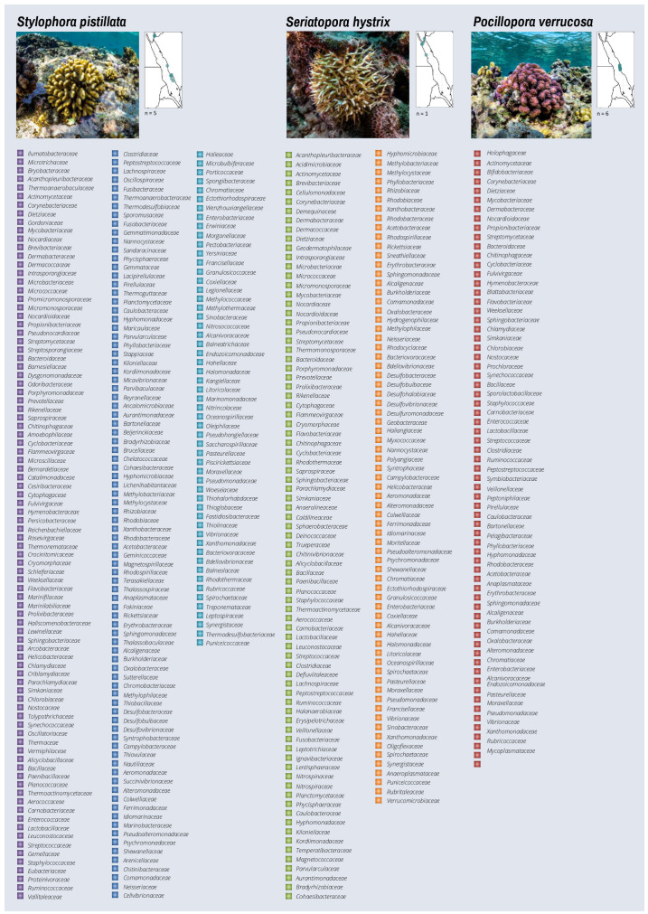
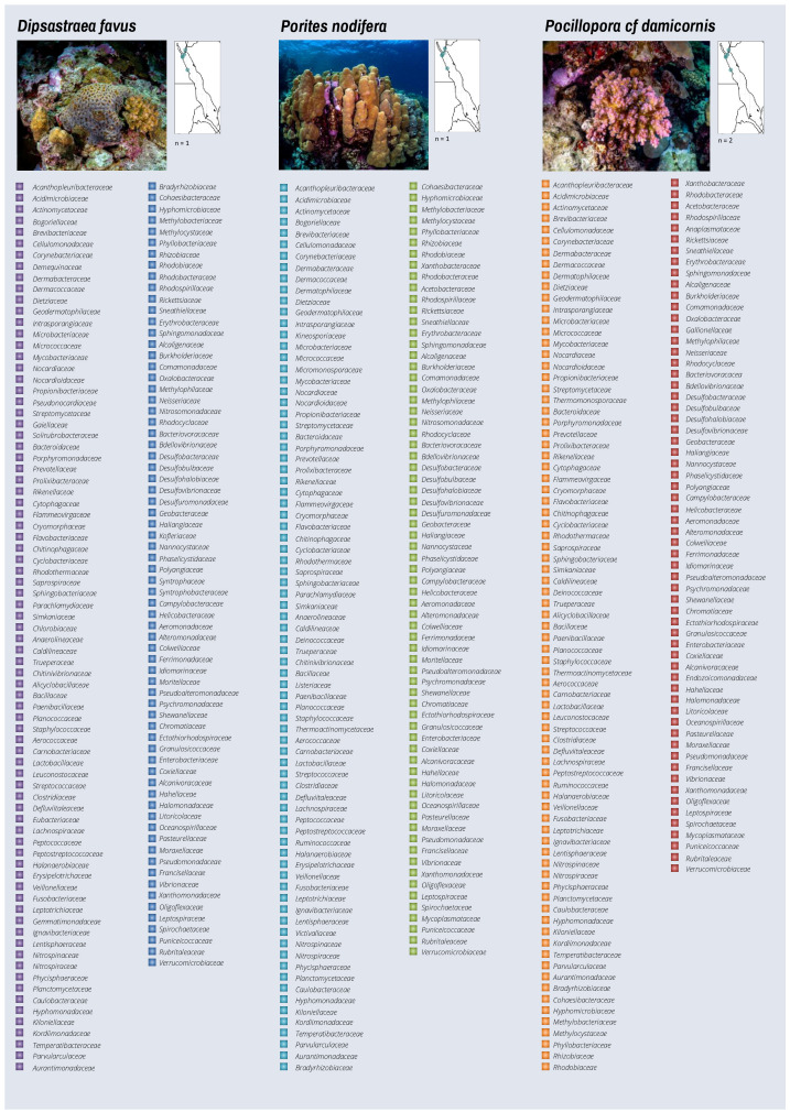
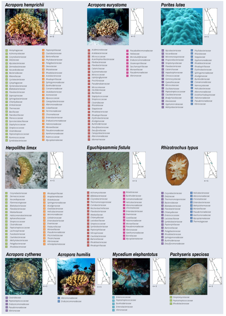
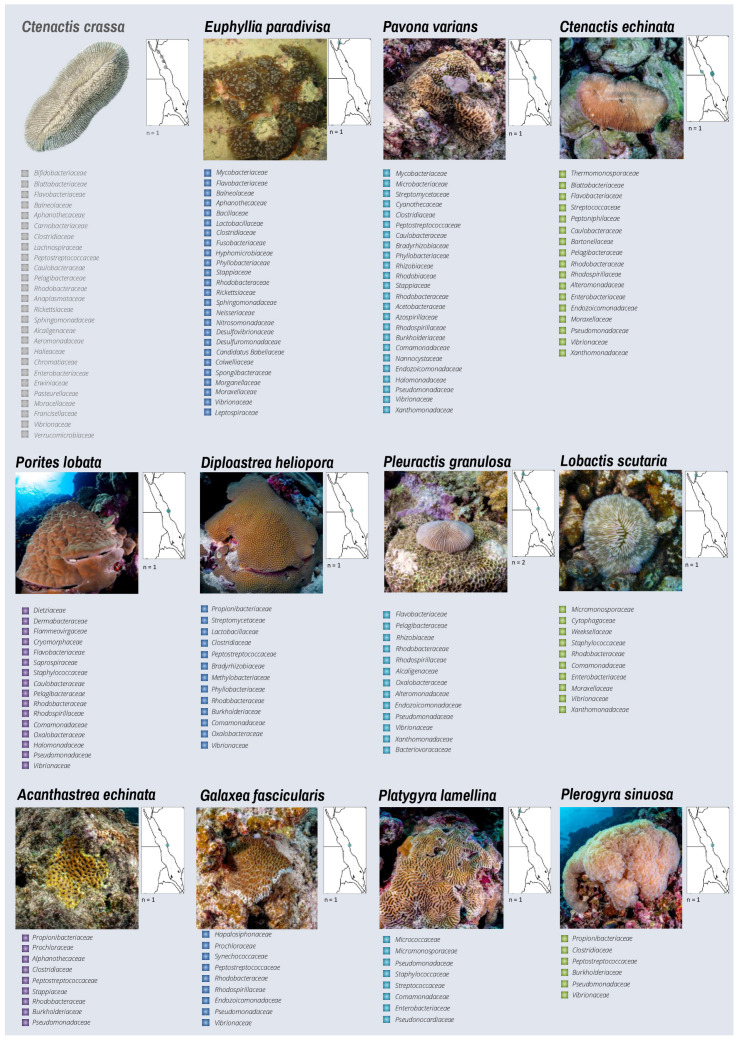
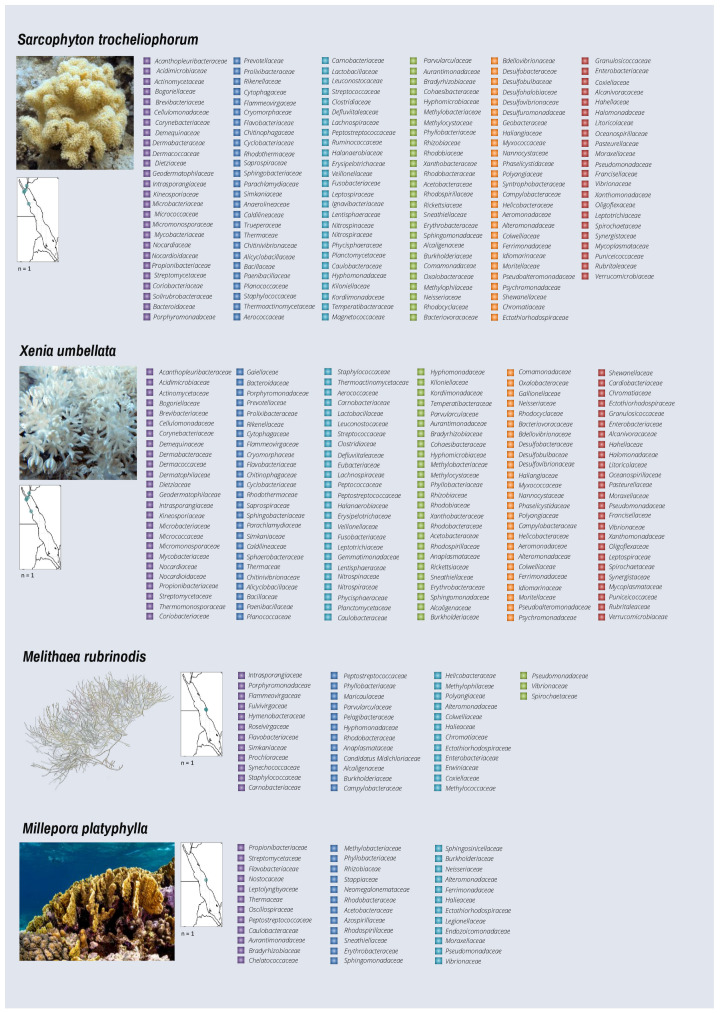
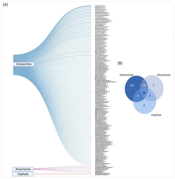
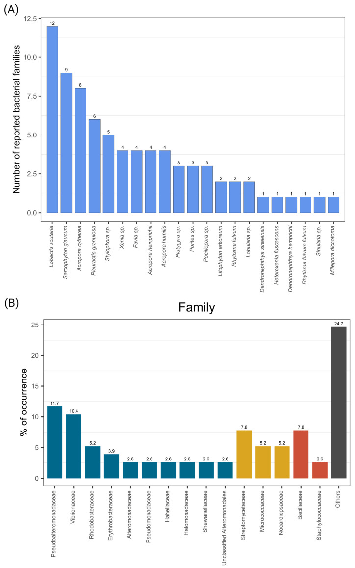
The Red Sea is a suitable model for studying coral reefs under climate change due to its strong environmental gradient that provides a window into future global warming scenarios. For instance, corals in the southern Red Sea thrive at temperatures predicted to occur at the end of the century in other biogeographic regions. Corals in the Red Sea thrive under contrasting thermal and environmental regimes along their latitudinal gradient. Because microbial communities associated with corals contribute to host physiology, we conducted a systematic review of the known diversity of Red Sea coral-associated bacteria, considering geographic location and host species. Our assessment comprises 54 studies of 67 coral host species employing cultivation-dependent and cultivation-independent techniques. Most studies have been conducted in the central and northern Red Sea, while the southern and western regions remain largely unexplored. Our data also show that, despite the high diversity of corals in the Red Sea, the most studied corals were Pocillopora verrucosa, Dipsastraea spp., Pleuractis granulosa, and Stylophora pistillata. Microbial diversity was dominated by bacteria from the class Gammaproteobacteria, while the most frequently occurring bacterial families included Rhodobacteraceae and Vibrionaceae. We also identified bacterial families exclusively associated with each of the studied coral orders: Scleractinia (n = 125), Alcyonacea (n = 7), and Capitata (n = 2). This review encompasses 20 years of research in the Red Sea, providing a baseline compendium for coral-associated bacterial diversity.
Keywords: Red Sea; bacterial diversity; coral reefs; coral-associated bacteria; microbial ecology; microbiology.
Conflict of interest statement
The authors declare that the research was conducted in the absence of any commercial or financial relationships that could be construed as a potential conflict of interest.
Figures
Similar articles
-
Disparate population and holobiont structure of pocilloporid corals across the Red Sea gradient demonstrate species-specific evolutionary trajectories.Mol Ecol. 2023 May;32(9):2151-2173. doi: 10.1111/mec.16871. Epub 2023 Mar 3. Mol Ecol. 2023. PMID: 36869609
-
Spatio-temporal analyses of Symbiodinium physiology of the coral Pocillopora verrucosa along large-scale nutrient and temperature gradients in the Red Sea.PLoS One. 2014 Aug 19;9(8):e103179. doi: 10.1371/journal.pone.0103179. eCollection 2014. PLoS One. 2014. PMID: 25137123 Free PMC article.
-
Hydroids (Cnidaria, Hydrozoa) from Mauritanian Coral Mounds.Zootaxa. 2020 Nov 16;4878(3):zootaxa.4878.3.2. doi: 10.11646/zootaxa.4878.3.2. Zootaxa. 2020. PMID: 33311142
-
Microbial diseases of corals and global warming.Environ Microbiol. 2002 Jun;4(6):318-26. doi: 10.1046/j.1462-2920.2002.00302.x. Environ Microbiol. 2002. PMID: 12071977 Review.
-
Building living systematic reviews and reporting standards for comparative microscopic analysis of white diseases in hard corals.Ecol Evol. 2024 Jul 4;14(7):e11616. doi: 10.1002/ece3.11616. eCollection 2024 Jul. Ecol Evol. 2024. PMID: 38975266 Free PMC article. Review.
Cited by
-
Metagenomic Characterization of Microbiome Taxa Associated with Coral Reef Communities in North Area of Tabuk Region, Saudia Arabia.Life (Basel). 2025 Mar 7;15(3):423. doi: 10.3390/life15030423. Life (Basel). 2025. PMID: 40141768 Free PMC article.
-
Sediment exposure decreases diversity in the surface mucus layer microbiome of Porites lobata at Honoli'i, Hawai'i.Front Microbiol. 2025 Jul 28;16:1626064. doi: 10.3389/fmicb.2025.1626064. eCollection 2025. Front Microbiol. 2025. PMID: 40792269 Free PMC article.
-
Probiotics reshape the coral microbiome in situ without detectable off-target effects in the surrounding environment.Commun Biol. 2024 Apr 9;7(1):434. doi: 10.1038/s42003-024-06135-3. Commun Biol. 2024. PMID: 38594357 Free PMC article.
-
Endozoicomonas dominance and Vibrionaceae stability underpin resilience in urban coral Madracis auretenra.PeerJ. 2025 Apr 15;13:e19226. doi: 10.7717/peerj.19226. eCollection 2025. PeerJ. 2025. PMID: 40256745 Free PMC article.
-
Heatwave-driven persistent microbes threaten the resilience of Mediterranean coral holobionts.Environ Microbiome. 2025 Aug 21;20(1):107. doi: 10.1186/s40793-025-00765-8. Environ Microbiome. 2025. PMID: 40842040 Free PMC article.
References
-
- Rohwer F., Seguritan V., Azam F., Knowlton N. Diversity and distribution of coral-associated bacteria. Mar. Ecol. Prog. Ser. 2002;243:1–10. doi: 10.3354/meps243001. - DOI
Publication types
Grants and funding
LinkOut - more resources
Full Text Sources
Miscellaneous
