Review
. 2022 Nov 29;10(12):2356.
doi: 10.3390/microorganisms10122356.

CD36-A Host Receptor Necessary for Malaria Parasites to Establish and Maintain Infection

Affiliations
Review

CD36-A Host Receptor Necessary for Malaria Parasites to Establish and Maintain Infection

Anna Bachmann et al. Microorganisms. .

Abstract

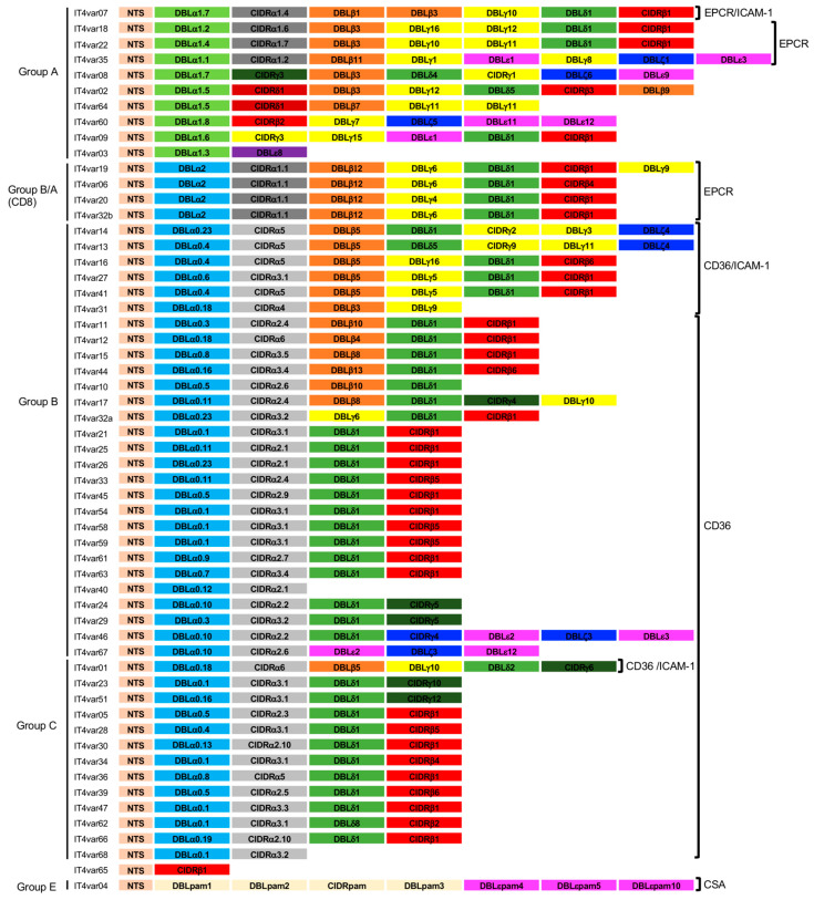
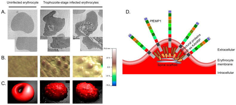
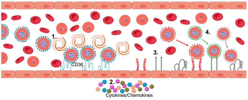
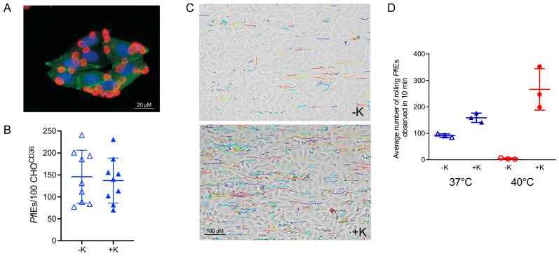
Plasmodium falciparum-infected erythrocytes (PfIEs) present P. falciparum erythrocyte membrane protein 1 proteins (PfEMP1s) on the cell surface, via which they cytoadhere to various endothelial cell receptors (ECRs) on the walls of human blood vessels. This prevents the parasite from passing through the spleen, which would lead to its elimination. Each P. falciparum isolate has about 60 different PfEMP1s acting as ligands, and at least 24 ECRs have been identified as interaction partners. Interestingly, in every parasite genome sequenced to date, at least 75% of the encoded PfEMP1s have a binding domain for the scavenger receptor CD36 widely distributed on host endothelial cells and many other cell types. Here, we discuss why the interaction between PfIEs and CD36 is optimal to maintain a finely regulated equilibrium that allows the parasite to multiply and spread while causing minimal harm to the host in most infections.

Keywords: CD36; Plasmodium falciparum; cytoadhesion; endothelial cell receptor; malaria; sequestration.


Conflict of interest statement

The authors declare no conflict of interest.

Figures

Figure 1
Figure 1
PfEMP1 repertoire of the P. falciparum isolate IT4, adapted from [17]. ECR binding phenotype [36]. Color code: light brown: N-terminal segment (NTS); bright green: Duffy binding-like (DBL)α1; light blue: DBLα2, DBLα0; dark grey: Cys rich inter-domain regions (CIDR)α1; light grey: CIDRα2–6; dark green: CIDRγ; orange: DBLβ; yellow: DBLγ; green: DBLδ; pink: DBLε; blue: DBLζ; purple: DBLε; IT4var04: light yellow: DBL/CIDRpam: pink: DBLεpam.
Figure 2
Figure 2
The knobs of PfIEs. (A) Transmission electron micrographs of uninfected and synchronised trophozoite-stage P. falciparum culture 24–28 h post-infection. The parasites were cultivated in the presence of human serum (10%), and the PfIEs were subjected to gelatin sedimentation to enrich knobby PfIEs [40,48]. (B,C) atomic force microscopic three-dimensional images of the surface of uninfected and trophozoite-stage PfIEs [40]. Images in (B) show magnifications directly from the membrane surface of the erythrocytes shown in (C). (D) Simplified schematic illustration of the structure of knobs.
Figure 3
Figure 3
Presumed sequence of sequestration of PfIEs to the vascular endothelium. 1. Adhesion and rolling over CD36. 2. Over time: endothelial activation, cytokine release. 3. Cytokine/chemokine-induced presentation of various receptors (e.g., ICAM-1, P-selectin, CD9). 4. Adhesion of PfEMP1s with different binding phenotypes (created with BioRender).
Figure 4
Figure 4
Cytoadhesion of knobby and knobless PfIEs to transgenic CHO cells presenting CD36 on the cell surface (CHOCD36) under static and flow conditions and at different temperatures. (A) Adhesion of PfIEs (red; anti-glycophorin A) to CHOCD36 cells (blue: nucleus (DAPI), green: cell surface (CD36-GFP fusion protein) under static binding conditions. (B) Cytoadhesion of knobbless (−K) and knobby (+K) PfIEs to CHOCD36 cells. (C) Trajectories showing the rolling binding behavior of knobless (−K) and knobby (+K) PfIEs to CHOCD36 cells. (D) Average number of knobless (−K) and knobby (+K) PfIEs adhering to CHOCD36 cells at 37 °C (blue) and 40 °C (red) at a shear stress of 0.9 dyn/cm2 [127].

References

    1. WHO . World Malaria Report 2021. WHO; Geneva, Switzerland: 2021.
    1. Saul A. The role of variant surface antigens on malaria-infected red blood cells. Parasitol. Today. 1999;15:455–457. doi: 10.1016/S0169-4758(99)01534-3. - DOI - PubMed
    1. Phillips M.A., Burrows J.N., Manyando C., van Huijsduijnen R.H., Van Voorhis W.C., Wells T.N.C. Malaria. Nat. Rev. Dis. Prim. 2017;3:17050. doi: 10.1038/nrdp.2017.50. - DOI - PubMed
    1. Craig A.G., Khairul M.F., Patil P.R. Cytoadherence and severe malaria. Malays. J. Med. Sci. 2012;19:5–18. - PMC - PubMed
    1. Newbold C., Craig A., Kyes S., Rowe A., Fernandez-Reyes D., Fagan T. Cytoadherence, pathogenesis and the infected red cell surface in Plasmodium falciparum. Int. J. Parasitol. 1999;29:927–937. doi: 10.1016/S0020-7519(99)00049-1. - DOI - PubMed