. 2022 Nov 30;10(12):2381.
doi: 10.3390/microorganisms10122381.

Generation and Utilization of a Monoclonal Antibody against Hepatitis B Virus Core Protein for a Comprehensive Interactome Analysis

Affiliations

Generation and Utilization of a Monoclonal Antibody against Hepatitis B Virus Core Protein for a Comprehensive Interactome Analysis

Yusuke Nakai et al. Microorganisms. .

Abstract

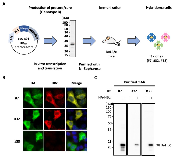
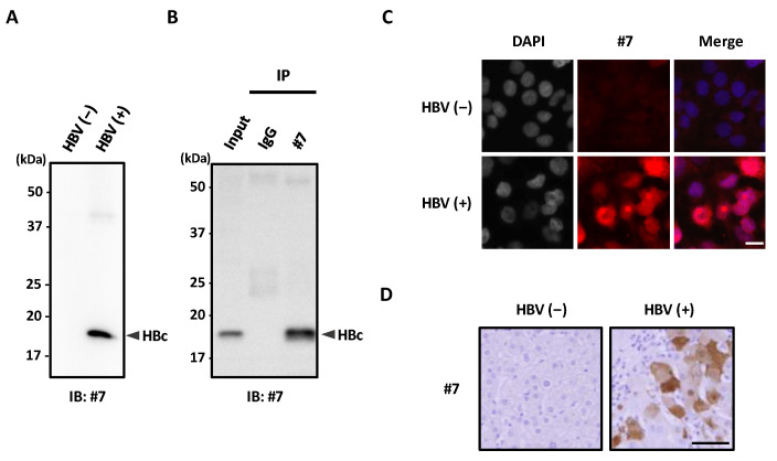
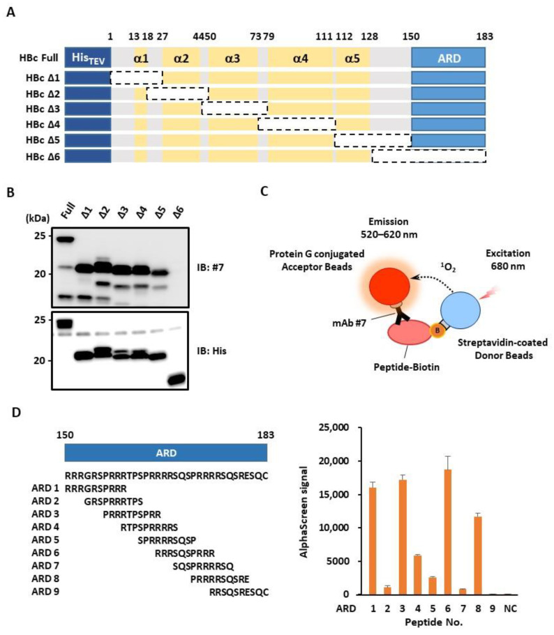
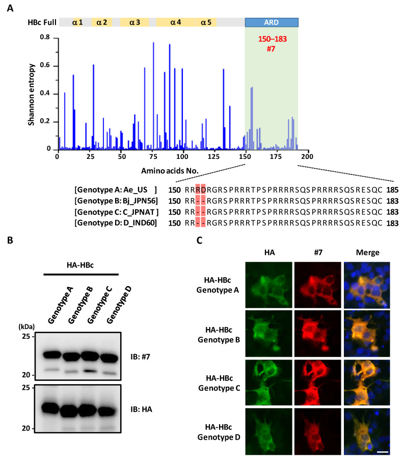
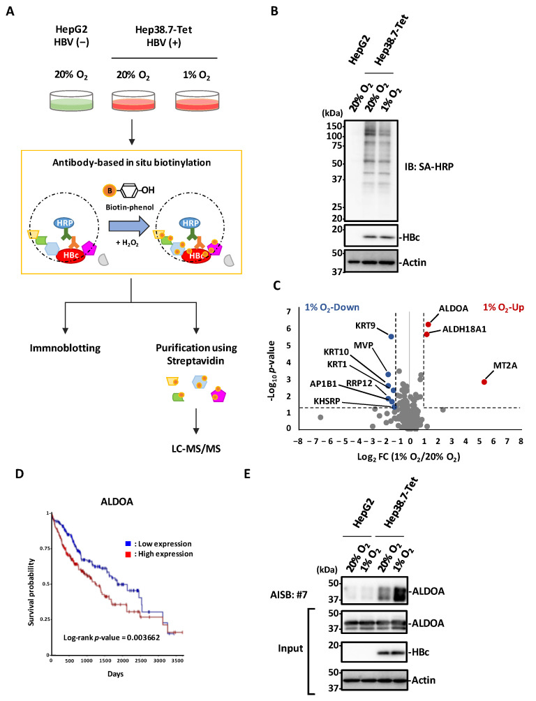
Hepatitis B virus (HBV) core antigen (HBc) is a structural protein that forms the viral nucleocapsid and is involved in various steps of the viral replication cycle, but its role in the pathogenesis of HBV infection is still elusive. In this study, we generated a mouse monoclonal antibody (mAb) against HBc and used it in antibody-based in situ biotinylation analysis in order to identify host proteins that interact with HBc. HBc antigen was produced with a wheat germ cell-free protein synthesis system and used to immunize mice. Among the established hybridoma clones, a single clone (mAb #7) was selected and further characterized for its ability in the antibody-based in situ biotinylation analysis to collect host proteins that are in the vicinity of HBc. Using mass spectrometry, we identified 215 HBc-interacting host proteins, three of which bind HBc most significantly under hypoxic conditions. Our results indicate that mAb #7 can be used to systematically identify host proteins that interact with HBc under pathophysiological conditions, and thus may be useful to explore the molecular pathways involved in HBV-induced cytopathogenesis.

Keywords: Hepatitis B virus; antibody-based in situ biotinylation; hypoxia; interactome analysis; monoclonal antibody.


Conflict of interest statement

Y.Y. is a current employee of Kanto Chemical Co., Inc. A.R. received a collaborative research grant from Kanto Chemical Co., Inc.

Figures

Figure 1
Figure 1
Production and screening of anti-HBc mouse mAbs. (A) Schematic of the generation of hybridoma cells that produce anti-HBc mouse mAbs. Recombinant His-precore/core (derived from HBV genotype B) was synthesized by a wheat germ cell-free system and purified with Ni-Sepharose. The red dot indicates the target protein. The purified protein was injected into BALB/c mice. Four weeks later, isolated lymphocytes were fused with myeloma cells, and hybridoma clones were established. SP6, SP6 promoter sequence; E01, translation enhancer sequence; His, histidine-tagged sequence; TEV, TEV protease-recognized sequence. (B,C) The reactivity of three mAbs (#7, #32, and #38) for HBc. HepG2 cells transfected with HA-HBc were fixed with 4% PFA and stained with the indicated mAbs (hybridoma supernatants, red; HA-antibody, green) and DAPI (blue). Scale bar, 10 μm (B). HepG2 cell lysates transfected with HA-HBc were analyzed by immunoblotting with the indicated mAbs (C).
Figure 2
Figure 2
The newly developed mAb #7 detects HBc in HBV-infected cells. (A) Lysates of HepG2 cells (HBV (−)) or HepG2.2.15.7 cells (HBV (+)) were analyzed by immunoblotting with mAb #7. (B) HepG2.2.15.7 cell lysates were immunoprecipitated with mAb #7, then bound proteins were analyzed by immunoblotting with mAb #7. (C) HepG2 cells (HBV (−)) and HepG2.2.15.7 cells (HBV (+)) were fixed with 4% PFA and then stained with mAb #7 (red) and DAPI (blue). Scale bar, 10 μm. (D) HBV negative (−) and positive (+) paraffin-embedded human liver tissues were stained with mAb #7 using peroxidase conjugate and DAB chromogen, and then counterstained with hematoxylin. Scale bar, 100 μm.
Figure 3
Figure 3
Epitope mapping for the newly developed mAb #7. (A) Schematic of a full-length HBc and six deletion mutants (HBcΔ1–6). These N-terminal His-tagged proteins were produced using a wheat germ cell-free system. ARD, arginine-rich domain. (B) His-HBc and its deletion mutants were analyzed by immunoblotting using mAb #7 or anti-His antibody. (C) Schematic of the AlphaScreen assay. Protein G-conjugated acceptor beads and streptavidin-conjugated donors were used to monitor the interaction of mAb #7 with the synthesized peptides. (D) AlphaScreen assay. Nine biotin-tagged ARD peptides (ARD1–9) were synthesized (left panel). The binding activity was measured as the level of the AlphaScreen luminescence signal (right panel). Error bars represent standard deviations from three independent experiments. NC, negative control.
Figure 4
Figure 4
mAb #7 detects HBc derived from multiple HBV genotypes. (A) Shannon entropy was calculated for each amino acid residue of HBc derived from 13,893 strains (upper panel). The mAb #7 binding region is represented in green. Multiple alignment of sequences in HBc ARD derived from genotypes Ae_US, Bj_JPN56, C_JPNAT, and D_IND60 (lower panel). The substituted amino acids are highlighted in red. (B) Recombinant HA-HBc (HBV genotypes A, B, C, and D) were synthesized using a wheat germ cell-free system and analyzed by immunoblotting with mAb #7 or anti-HA antibody. (C) HepG2 cells transfected with HA-HBc (HBV genotypes A, B, C, and D) were fixed with 4% PFA and stained with mAb #7 (red) or anti-HA antibody (green), and then counterstained with DAPI (blue). Scale bar, 10 μm.
Figure 5
Figure 5
Antibody-based in situ biotinylation analysis with mAb #7. (A) Schematic of proteomic analysis for the identification of host proteins that interact with HBc in HBV-positive (Hep38.7-Tet) and HBV-negative (HepG2) cells under normoxic and hypoxic conditions. Each cell was fixed and permeabilized. After mAb #7 bound to HBc, the proximal portion of HBc was biotinylated by adding biotin-phenol and H2O2. The cell extracts were analyzed by immunoblotting, followed by mass spectrometric analysis of the biotinylated proteins. (B) Biotinylation of each cell extract was confirmed by immunoblot analysis with Streptavidin HRP Conjugate. (C) Volcano plot representing differences in abundance of 215 host proteins near HBc under normoxic and hypoxic conditions. The x-axis indicates the log2 fold change and the y-axis indicates the –log10 p-value based on the two-tailed Student’s t-test. The proteins that were significantly upregulated (red) or downregulated (blue) under the hypoxic condition were identified based on two criteria: |fold change| > 2 and p-value < 0.05. The dotted lines show the criteria. FC, fold change. (D) Kaplan-Meier analysis of ALDOA was performed using the UCSC Xena platform and mRNA sequence databases of the Cancer Genome Atlas Program (TCGA). Log-rank p = 0.003662. Log-rank p-value < 0.05 was regarded as statistically significant. The x-axis indicates the days and the y-axis indicates the survival probability. (E) HBc interacts with ALDOA. The streptavidin-purified biotinylated samples were analyzed by immunoblotting with anti-ALDOA antibody. AISB, antibody-based in situ biotinylation.

Similar articles

Cited by

References

    1. Megahed F.A.K., Zhou X., Sun P. The interactions between HBV and the innate immunity of hepatocytes. Viruses. 2020;12:285. doi: 10.3390/v12030285. - DOI - PMC - PubMed
    1. D’Arienzo V., Ferguson J., Giraud G., Chapus F., Harris J.M., Wing P.A.C., Claydon A., Begum S., Zhuang X., Balfe P., et al. The CCCTC-binding factor CTCF represses Hepatitis B virus enhancer I and regulates viral transcription. Cell. Microbiol. 2021;23:e13274. doi: 10.1111/cmi.13274. - DOI - PMC - PubMed
    1. Virzi A., Motos V.G., Tripon S., Baumert T.F., Virzi A., Motos V.G., Tripon S., Baumert T.F., Profi- J.L., Virz A., et al. Profibrotic Signaling and HCC Risk during Chronic Viral Hepatitis: Biomarker Development To cite this version: HAL Id: Hal-03603544 Profibrotic Signaling and HCC Risk during Chronic Viral Hepatitis: Biomarker Development. J. Clin. Med. 2021;10:977. - PMC - PubMed
    1. Fisman D.N., Agrawal D., Leder K. The effect of age on immunologic response to recombinant hepatitis B vaccine: A meta-analysis. Clin. Infect. Dis. 2002;35:1368–1375. doi: 10.1086/344271. - DOI - PubMed
    1. Pattyn J., Hendrickx G., Vorsters A., Van Damme P. Hepatitis B Vaccines. J. Infect. Dis. 2021;224((Suppl. 4)):S343–S351. doi: 10.1093/infdis/jiaa668. - DOI - PMC - PubMed

LinkOut - more resources