. 2022 Dec 7;27(24):8639.
doi: 10.3390/molecules27248639.

Opuntia dillenii Haw. Polysaccharide Promotes Cholesterol Efflux in THP-1-Derived Foam Cells via the PPARγ-LXRα Signaling Pathway

Affiliations

Opuntia dillenii Haw. Polysaccharide Promotes Cholesterol Efflux in THP-1-Derived Foam Cells via the PPARγ-LXRα Signaling Pathway

Heng Li et al. Molecules. .

Abstract

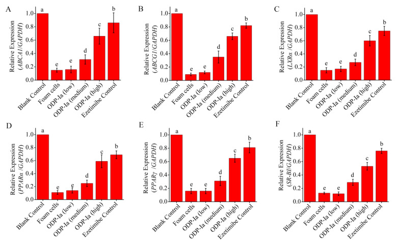
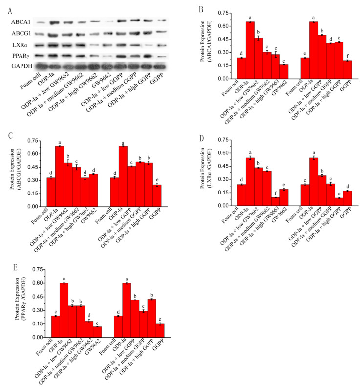
There is increasing evidence supporting a role for enhanced macrophage cholesterol efflux in ameliorating atherosclerosis. Opuntia dillenii Haw. polysaccharide (ODP-Ia), the most important functional component obtained from Opuntia dillenii Haw. stem, has anti-atherosclerosis effects. Therefore, we propose that ODP-Ia could promote cholesterol efflux via the PPARγ-LXRα signaling pathway. In this study, THP-1 foam cells derived from macrophages were treated with different concentrations of ODP-Ia, GGPP (antagonist of LXRα) and GW9662 (antagonist of PPARγ), with or without 15 nmol ODP-Ia. The total cholesterol content in the cells was measured. The mRNA of ABCA1, ABCG1, PPARγ, LXRα and their protein levels in the foam cells were detected by RT−PCR and Western blot, respectively. The results showed that ODP-Ia plays a role in significantly promoting cholesterol efflux (p < 0.05) by upregulating the expression of ABCA1, ABCG1, SR-BI, PPARγ, PPARα and LXRα. Meanwhile, PPARγ and LXRα antagonists dramatically interfered the cholesterol efflux mediated by ODP-Ia (p < 0.05) and dramatically inhibited the upregulating effect of ODP-Ia on the expression of PPARγ, LXRα, ABCA1 and ABCG1 at both protein and mRNA levels (p < 0.05). In conclusion, ODP-Ia promotes cholesterol efflux in the foam cells through activating the PPARγ-LXRα signaling pathway. This bioactivity suggested that ODP-Ia may be of benefit in treating atherosclerosis.

Keywords: ABCA1; Opuntia dillenii Haw. polysaccharide; PPARγ-LXRα; THP-1-derived foam cells; cholesterol efflux.


Conflict of interest statement

The authors declare that the research was conducted in the absence of any commercial or financial relationships that could be construed as a potential conflict of interest.

Figures

Figure 1
Figure 1
Macrophages and Oil red O staining maps of foam cells (Magnification, ×100). (A) After 24 h of PMA stimulation, the cells differentiated into macrophages. (B) After 48 h of incubation with 50 ng/mL ox-LDL, the macrophages developed into foam cells, and intracellular lipid droplets were stained with Oil red O.
Figure 2
Figure 2
Effect of ODP-Ia and ezetimibe on THP-1 macrophage-derived foam cell viability, as determined through MTT analysis. Values are mean ± SD, n = 5. (A) ODP-Ia. (B) Ezetimibe. Different superscript letters indicate multiple comparisons with significant differences (SNK-q test, p < 0.05).
Figure 3
Figure 3
Effects of apoA-I on cholesterol accumulation in THP-1 macrophage-derived foam cells. Values are mean ± SD, n = 5. (A,B) Total cholesterol content of THP-1 macrophages decreased in a time- and concentration-dependent manner in the apoA-I-treated foam cells. (C) apoA-I-induced cholesterol efflux mediated by ODP-Ia. Different superscript letters indicate multiple comparisons with significant differences (SNK-q test, p < 0.05).
Figure 4
Figure 4
Effects of ODP-Ia on the cholesterol outflow in foam cells. (mean ± SD, n = 5). Different superscript letters indicate multiple comparisons with significant differences (SNK-q test, p < 0.05).
Figure 5
Figure 5
mRNA expression of cholesterol efflux-associated gene mediated by ODP-Ia. Values are mean ± SD, n = 9. (A) ABCA1 mRNA. (B) ABCG1 mRNA. (C) LXRα mRNA. (D) PPARα mRNA. (E) PPARγ mRNA. (F) SR-BI mRNA. Different superscript letters indicate multiple comparisons with significant differences (SNK-q test, p < 0.05).
Figure 6
Figure 6
Effect of ODP-Ia on protein expression of ABCA1, ABCG1, LXRα, PPARα, PPARγ and SR-BI in THP-1 macrophage-derived foam cells. Dates in figure represent the density values of each band (mean ± SD, n = 9). (A) ABCA1 protein expression level. (B) ABCG1 protein expression level. (C) LXRα protein expression level. (D) PPARα protein expression level. (E) PPARγ protein expression level. (F) SR-BI protein expression level. Different superscript letters indicate multiple comparisons with significant differences (SNK-q test, p < 0.05).
Figure 7
Figure 7
The ODP-Ia-induced cholesterol efflux mediated by GGPP (A) and GW9662 (B) (mean ± SD, n = 5). Different superscript letters indicate multiple comparisons with significant differences (SNK-q test, p < 0.05).
Figure 8
Figure 8
mRNA expression of cholesterol efflux associated gene mediated by GGPP and ODP-Ia. (mean ± SD, n = 3). (A) ABCA1 mRNA. (B) ABCG1 mRNA. (C) LXRα mRNA. (D) PPARγ mRNA. Different superscript letters indicate multiple comparisons with significant differences (SNK-q test, p < 0.05).
Figure 9
Figure 9
mRNA expression of cholesterol efflux associated gene mediated by GW9662 and ODP-Ia. (mean ± SD, n = 3). (A) ABCA1 mRNA. (B) ABCG1 mRNA. (C) LXRα mRNA. (D) PPARγ mRNA. Different superscript letters indicate multiple comparisons with significant differences (SNK-q test, p < 0.05).
Figure 10
Figure 10
Protein expression of cholesterol efflux-associated gene mediated by GW9662 and GGPP. Dates in figure represent the density values of each band (mean ± SD, n = 9) (A) Western blot analyses of ABCA1, ABCG1, LXRα and PPARγ in THP-1-derived foam cells. (B) ABCA1 protein expression level. (C) ABCG1 protein expression level. (D) LXRα protein expression level. (E) PPARγ protein expression level. Different superscript letters indicate multiple comparisons with significant differences (SNK-q test, p < 0.05).

Similar articles

Cited by

References

    1. Xu Y., Liu C., Han X., Jia X., Li Y., Liu C., Li N., Liu L., Liu P., Jiang X., et al. E17241 as a Novel ABCA1 (ATP-Binding Cassette Transporter A1) Upregulator Ameliorates Atherosclerosis in Mice. Arter. Thromb. Vasc. Biol. 2021;41:e284–e298. doi: 10.1161/ATVBAHA.120.314156. - DOI - PubMed
    1. Brown M.S., Goldstein J.L. Lipoprotein Metabolism in the Macrophage: Implications for Cholesterol Deposition in Atherosclerosis. Annu. Rev. Biochem. 1983;52:223–261. doi: 10.1146/annurev.bi.52.070183.001255. - DOI - PubMed
    1. Guo L., Jiang B., Li D., Xiao X. Nephroprotective Effect of Adropinin against Streptozotocin-Induced Diabetic Nephropathy in Rats: Inflammatory Mechanism and YAP/TAZ Factor. Drug Des. Dev. Ther. 2021;15:589–600. doi: 10.2147/DDDT.S294009. - DOI - PMC - PubMed
    1. Nasser M., Zhu S., Huang H., Zhao M., Wang B., Ping H., Geng Q., Zhu P. Macrophages: First guards in the prevention of cardiovascular diseases. Life Sci. 2020;250:117559. doi: 10.1016/j.lfs.2020.117559. - DOI - PubMed
    1. Zou J., Xu C., Zhao Z.-W., Yin S.-H., Wang G. Asprosin inhibits macrophage lipid accumulation and reduces atherosclerotic burden by up-regulating ABCA1 and ABCG1 expression via the p38/Elk-1 pathway. J. Transl. Med. 2022;20:1–16. doi: 10.1186/s12967-022-03542-0. - DOI - PMC - PubMed

MeSH terms