. 2022 Dec 8;14(24):5233.
doi: 10.3390/nu14245233.

Heat-Killed Bifidobacterium bifidum B1628 May Alleviate Dextran Sulfate Sodium-Induced Colitis in Mice, and the Anti-Inflammatory Effect Is Associated with Gut Microbiota Modulation

Affiliations

Heat-Killed Bifidobacterium bifidum B1628 May Alleviate Dextran Sulfate Sodium-Induced Colitis in Mice, and the Anti-Inflammatory Effect Is Associated with Gut Microbiota Modulation

Cuijiao Feng et al. Nutrients. .

Abstract

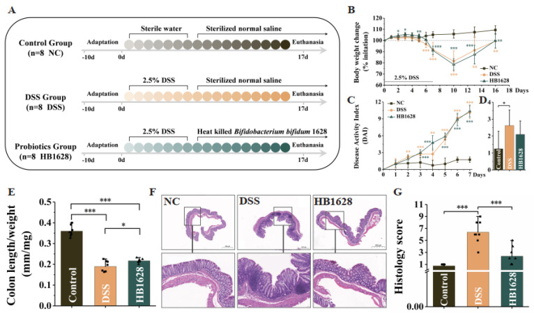
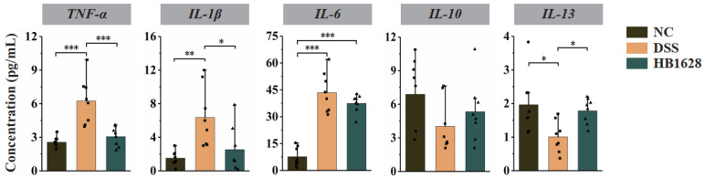
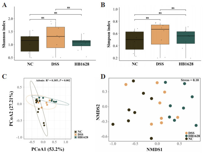
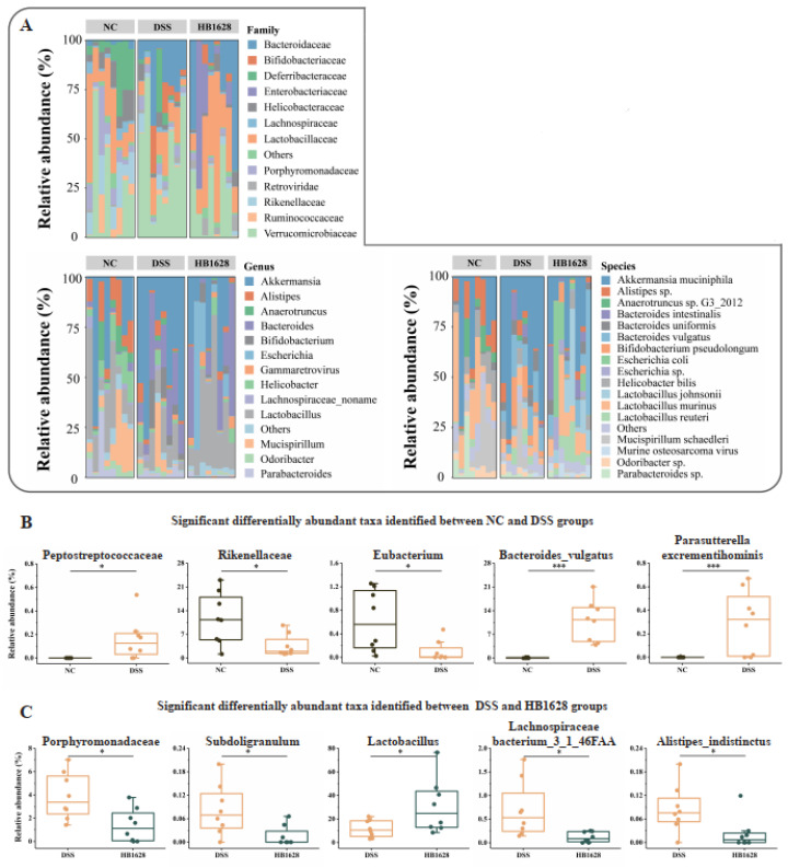
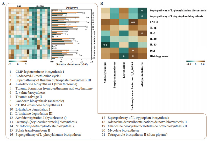
Inflammatory bowel disease (IBD) is a chronic inflammatory disease associated with gut dysbiosis. This study aimed to investigate the effects of heat-killed Bifidobacterium bifidum B1628 (HB1628) in dextran sulfate sodium (DSS)-induced colitis in mice. The following three mouse groups were included (n = eight per group): NC (normal control), DSS (colitis), and HB1628 (colitis and postbiotic). The mice in the DSS group showed significant weight loss and histological damage, developed bloody diarrhea, scored high in the disease activity index (DAI), and exhibited increases in pro-inflammatory cytokines (interleukin [IL]-1β, IL-6, and tumor necrosis factor [TNF]-α) and decreases in an anti-inflammatory cytokine (IL-13) in the serum. These changes were accompanied by gut microbiota modulation in colitis mice (decreases in Rikenellaceae and Eubacterium; increases in Peptostreptococcaceae, Bacteroides vulgatus, and Parasutterella excrementihominis). The HB1628 group had lower DAIs, histology scores, and serum levels of pro-inflammatory cytokines (IL-1β and TNF-α), but higher levels of an anti-inflammatory cytokine (IL-13), compared with the DSS group, suggesting a less severe inflammatory state after the HB1628 intervention. Additionally, HB1628 improved DSS-induced gut dysbiosis, which is evidenced by increases in intestinal beneficial bacteria, such as Lactobacillus, and decreases in known unfavorable taxa in IBD, e.g., Porphyromonadaceae, Subdoligranulum, Lachnospiraceae bacterium 3_1_46FAA, and Alistipes indistinctus. Functional metagenomics revealed three significantly enriched metabolic pathways in the HB1628 group (namely, the aerobic respiration I [cytochrome c] pathway and the superpathways of L-phenylalanine biosynthesis and L-tryptophan biosynthesis, respectively). In conclusion, our results showed that HB1628 effectively improved the inflammation state and tissue damage in DSS-induced colitis mice, and the symptom relief effect was accompanied by obvious gut microbiota remodulation.

Keywords: Bifidobacterium bifidum; anti-inflammation; gut microbiota; heat-killed; inflammatory bowel disease; postbiotics.


Conflict of interest statement

The authors declare that they have no known competing financial interest or personal relationships that could have appeared to influence the work reported in this paper.

Figures

Figure 1
Figure 1
Organic acids and short-chain fatty acids in heat-killed Bifidobacterium bifidum B1628. The green gradient represents different kinds of organic acids, and the red gradient represents different kinds of short-chain fatty acids. The error bars represent standard deviations for the five determinations.
Figure 2
Figure 2
Heat-killed Bifidobacterium bifidum B1628 attenuated colonic damage in dextran sulfate sodium (DSS)-induced colitis. (A) Experimental design. (B) Daily body weight change. (C,D) Disease activity index during the DSS induction and at day 18. (E) Colon length/weight ratio. (F) Representative hematoxylin- and eosin-stained micrographs of colon tissue sections (at 20× and 200× magnification) at day 18. (G) Histology score. Control group (NC): without DSS induction; DSS group (DSS): 2.5% DSS induction; HB1628 group (HB1628): 2.5% DSS and 0.2 mL of Bifidobacterium bifidum B1628 (n = eight per group; * represents p < 0.05, ** represents p < 0.01, *** represents p < 0.001). The error bars represent the standard deviation.
Figure 3
Figure 3
Serum cytokine levels of the three mouse groups at day 18. The serum concentrations of five cytokines were determined, i.e., tumor necrosis factor-α (TNF-α), interleukin (IL)-1β, IL-6, IL-10, and IL-13. Control group (NC): without DSS induction; DSS group (DSS): 2.5% DSS induction; HB1628 group (HB1628): 2.5% DSS and 0.2 mL of Bifidobacterium bifidum B1628 (n = eight per group; * represents p < 0.05, ** represents p < 0.01, *** represents p < 0.001).
Figure 4
Figure 4
Diversity of gut microbiota in the three mouse groups at day 18. (A) Shannon index. (B) Simpson index. (C) Principal coordinate analysis (PCoA, Bray–Curtis distance) and (D) non-metric multidimensional scaling (NMDS, Bray–Curtis distance). Control group (NC): without DSS induction; DSS group (DSS): 2.5% DSS induction; HB1628 group (HB1628): 2.5% DSS and 0.2 mL of Bifidobacterium bifidum B1628 (n = eight per group; “ns” means not significant).
Figure 5
Figure 5
Taxonomic distribution of mouse gut microbiota and significant differentially abundant gut microbes. (A) Family-, genus-, and species-level distributions of the mouse gut microbiota. Significant differentially abundant gut bacterial families, genera, and species between the (B) NC and DSS groups and the (C) DSS and HB1628 groups. Control group (NC): without DSS induction; DSS group (DSS): 2.5% DSS induction; HB1628 group (HB1628): 2.5% DSS and 0.2 mL of Bifidobacterium bifidum B1628 (n = eight per group; * represents p < 0.05, *** represents p < 0.001).
Figure 6
Figure 6
Gut metagenomic potential and correlation analyses. (A) Significant differences in the gut metagenomic potentials between the DSS (colitis) and HB1628 (colitis and postbiotic intervention) groups (n = eight per group). The color scale represents the relative abundance of specific pathways encoded in the fecal metagenome (ranging from a high relative abundance [2; dark brown] to a low relative abundance [−2; dark green]). The error bars represent the standard deviation. (B) Correlation heat map of the Spearman’s rank correlation coefficient between the differentially abundant taxa and various parameters. The color scale represents the strength of the correlation coefficient (ranging from 0.6 [brown, a strong positive correlation] to −0.6 [green, a strong negative correlation]). * represents p < 0.05; ** represents p < 0.01. TNF = tumor necrosis factor; IL = interleukin; DAI = disease activity index.

References

    1. Agrawal M., Allin K.H., Petralia F., Colombel J.-F., Jess T. Multiomics to elucidate inflammatory bowel disease risk factors and pathways. Nat. Rev. Gastroenterol. Hepatol. 2022;19:399–409. doi: 10.1038/s41575-022-00593-y. - DOI - PMC - PubMed
    1. Turner D., Ricciuto A., Lewis A., D’Amico F., Dhaliwal J., Griffiths A.M., Bettenworth D., Sandborn W.J., Sands B.E., Reinisch W., et al. STRIDE-II: An Update on the Selecting Therapeutic Targets in Inflammatory Bowel Disease (STRIDE) Initiative of the International Organization for the Study of IBD (IOIBD): Determining Therapeutic Goals for Treat-to-Target strategies in IBD. Gastroenterology. 2021;160:1570–1583. doi: 10.1053/j.gastro.2020.12.031. - DOI - PubMed
    1. Teofani A., Marafini I., Laudisi F., Pietrucci D., Salvatori S., Unida V., Biocca S., Monteleone G., Desideri A. Intestinal taxa abundance and diversity in inflammatory bowel disease patients: An analysis including covariates and confounders. Nutrients. 2022;14:260. doi: 10.3390/nu14020260. - DOI - PMC - PubMed
    1. Lee M., Chang E.B. Inflammatory Bowel Diseases (IBD) and the Microbiome—Searching the Crime Scene for Clues. Gastroenterology. 2021;160:524–537. doi: 10.1053/j.gastro.2020.09.056. - DOI - PMC - PubMed
    1. Liu S., Zhao W., Lan P., Mou X. The microbiome in inflammatory bowel diseases: From pathogenesis to therapy. Protein Cell. 2021;12:331–345. doi: 10.1007/s13238-020-00745-3. - DOI - PMC - PubMed

LinkOut - more resources