. 2022 Dec 6;11(12):1479.
doi: 10.3390/pathogens11121479.

Host tRNA-Derived RNAs Target the 3'Untranslated Region of SARS-CoV-2

Affiliations

Host tRNA-Derived RNAs Target the 3'Untranslated Region of SARS-CoV-2

Emily N Hendrickson et al. Pathogens. .

Abstract

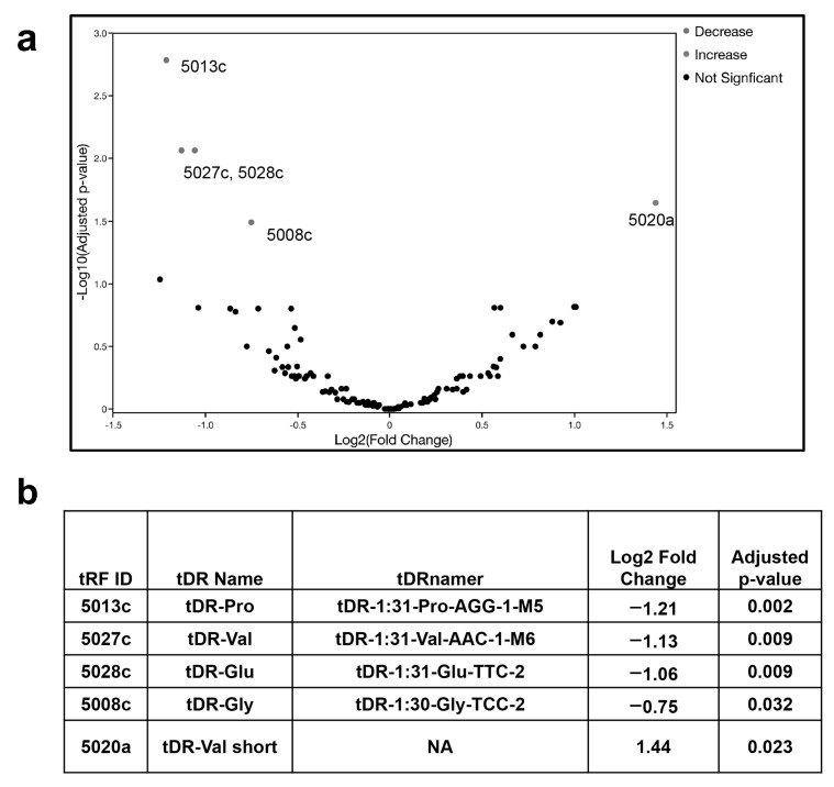
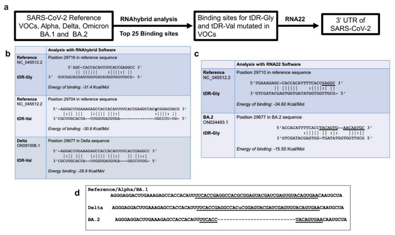
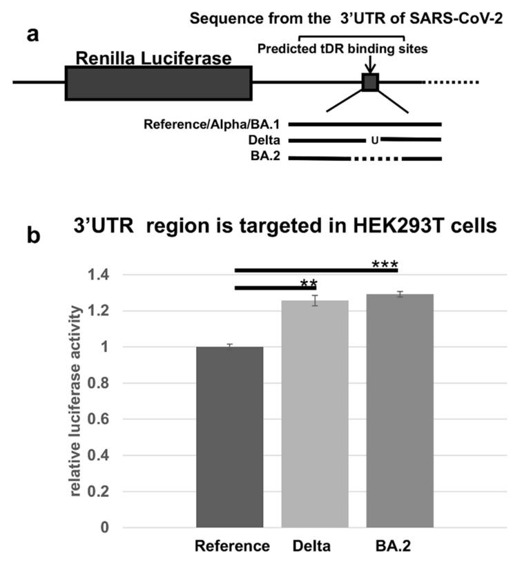
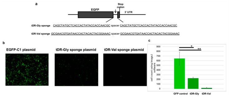
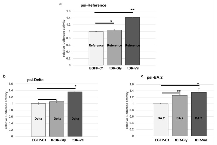
The COVID-19 pandemic revealed a need for new understanding of the mechanisms regulating host-pathogen interactions during viral infection. Transfer RNA-derived RNAs (tDRs), previously called transfer RNA fragments (tRFs), have recently emerged as potential regulators of viral pathogenesis. Many predictive studies using bioinformatic approaches have been conducted providing a repertoire of potential small RNA candidates for further analyses; however, few targets have been validated to directly bind to SARS-CoV-2 sequences. In this study, we used available data sets to identify host tDR expression altered in response to SARS-CoV-2 infection. RNA-interaction-prediction tools were used to identify sequences in the SARS-CoV-2 genome where tDRs could potentially bind. We then developed luciferase assays to confirm direct regulation through a predicted region of SARS-CoV-2 by tDRs. We found that two tDRs were downregulated in both clinical and in vitro cell culture studies of SARS-CoV-2 infection. Binding sites for these two tDRs were present in the 3' untranslated region (3'UTR) of the SARS-CoV-2 reference virus and both sites were altered in Variants of Concern (VOCs) that emerged later in the pandemic. These studies directly confirm the binding of human tDRs to a specific region of the 3'UTR of SARS-CoV-2 providing evidence for a novel mechanism for host-pathogen regulation.

Keywords: SARS-CoV-2; VOCs; Variants of Concern; host-viral interactions; small noncoding RNA; tDR; tRF; tRNA fragments; tRNA-derived RNAs.


Conflict of interest statement

The authors declare no conflict of interest.

Figures

Figure 1
Figure 1
Altered expression of a subset of human tDRs in plasma from SARS-CoV-2-infected patients compared to plasma samples from uninfected individuals. (a) Volcano plot showing the differential expression of tDRs in SARS-CoV-2-infected patients. The significantly altered tDRs are indicated by color with red being increased and blue designating the decreased tDRs, the p-value cut-off is <0.05. Log2 fold change, and p-value are those corresponding to the volcano plot. (b) Table of tDRs significantly altered in SARS-CoV-2-infected patients compared to uninfected individuals with the naming conventions: tRF ID is from the original data base used in the analysis and using the former name of tRNA fragments (http://genome.bioch.virginia.edu/trfdb/ [27,28] accessed on 17 January 2022); the tDR name is a shortened name used for convenience in this manuscript; the tDRnamer is the most accurate naming convention for the tRNA-derived RNAs [29,30], http://trna.ucsc.edu/tDRnamer/docs/ accessed on 12 November 2022; NA is not applicable for 5020a because it is too short to be identified in the tDRnamer data base (http://trna.ucsc.edu/tDRnamer/index.html) accessed on 12 November 2022.
Figure 2
Figure 2
Predicted binding of tDR-Gly and tDR-Val to the SARS-CoV-2 genome. (a) A schematic of the process used to identify potential hybridization sites for tDRs in the SARS-CoV-2 genomic sequences. RNAhybrid was used to predict the top 25 binding sites for tDR-Gly and tDR-Val in the SARS-CoV-2 reference sequence (NC_045512.2), and in the VOCs, Alpha (OV054768.1), Delta (OK091006.1), Omicron BA.1 (OL672836.1) and BA.2 (ON024493.1). The hybridization prediction software, RNA22, which uses a different algorithm than RNAhybrid was also used to test each tDR against the indicated sequences. The application of these two methods led to the choice of a site in the 3′UTR of SARS-CoV-2, for further analysis. (b) RNAhybrid predictions for tDR-Gly and tDR-Val binding to the 3′ end of SARS-CoV-2 showed a minimal free energy of binding similar to that expected for a microRNA and its target [33]. The sequence in this region of the Delta VOC contains a single nucleotide polymorphism changing a G in the reference sequence to a U in Delta. The alteration in the Delta VOC changes the predicted binding shown for tDR-Val to the Delta VOC. (c) The RNA22 prediction software for the 3′UTR of SARS-CoV-2 showed that tDR-Gly but not tDR-Val was predicted to bind, and binding was predicted to be altered in the BA.2 sequence. The sites predicted by RNAhybrid and RNA22 are slightly different due to the numbering conventions and parameters for each software package. (d) Sequences in the region of SARS-CoV-2 predicted to bind to tDR-Gly or Val are not altered in the Alpha or Omicron BA.1 variants. A base-pair change is detected in this region in the Delta VOC (u) and a 26-base-pair deletion is observed in the BA.2 variant. The underlined bases in the Reference/Alpha/Omicron BA.1 sequences designate the stem loop II (s2m) region of SARS-CoV-2 [34,35], which is partially deleted in BA.2.
Figure 3
Figure 3
Direct regulation through the 3′UTR of SARS-CoV-2 as detected by the loss of luciferase activity. (a) A schematic showing the luciferase constructs in the psi-Check-2 plasmid; the reference sequence is the same in the VOCs Alpha and Omicron BA.1 in this region; Delta is altered by one base and the sequence for BA.2 is the region of the 3′UTR encompassing a 26-base-pair deletion. (b) Human embryonic kidney cells (HEK293T) were transfected with the luciferase constructs. The transfections were performed in triplicate. The luciferase activity of both VOCs was compared to the reference sequence from SARS-CoV-2 as an unmutated control. The alterations in the sequence resulted in significant changes in the luciferase activity in Delta (** p-value < 0.0005) or in BA.2 (*** p-value < 0.00005). The transfections were performed in triplicate. The results were compared to the reference sequence as a percent of control and the error bars refer to ±standard deviation.
Figure 4
Figure 4
Plasmids with the anti-sense sponge sequences for tDR-Gly and tDR-Val were transfected into the HEK293T cells. (a) A schematic showing the sponge constructs in the EGFP-C1 plasmid. Two copies of the anti-sense sequence for tDR-Gly and tDR-Val were inserted at the 3′ end of the EGFP protein-encoding sequence. The anti-sense sequences for tDR-Gly and tDR-Val are underlined. (b) HEK293T cells were transfected with either the empty vector EGFP-C1 plasmid, tDR-Gly-EGFP-C1 sponge (tDR-Gly) or the tDR-Val-EGFP-C1 sponge (tDR-Val). (c) ImageJ software [38] was used to count the green cells in triplicate wells. The results were compared to the EGFP-C1 plasmid (GFP control). Error bars refer to ±standard deviation (* p-value < 0.05, ** p-value < 0.005).
Figure 5
Figure 5
Sponges specific to tDR-Gly and tDR-Val block the targeting of the 3′UTR sequences from SARS-CoV-2. (a) HEK293T cells were transfected with the psi-reference sequence and either the EGFP-C1 empty vector control (EGFP-C1), tDR-Gly EGFP-C1 sponge (tDR-Gly) or the tDR-Val EGFP-C1 sponge (tDR-Val). (b) HEK293T cells transfected as in (a) but with the psi-Delta construct. (c) HEK293T cells transfected as in (a) but with the psi-BA.2 construct. The transfections were performed in triplicate. The results were compared to the EGFP-C1 empty vector (control) as a percent of control and the error bars refer to ±standard deviation (* p-value ≤ 0.05, ** p-value < 0.005).

Similar articles

Cited by

References

    1. Zhu N., Zhang D., Wang W., Li X., Yang B., Song J., Zhao X., Huang B., Shi W., Lu R., et al. A Novel Coronavirus from Patients with Pneumonia in China, 2019. N. Engl. J. Med. 2020;382:727–733. doi: 10.1056/NEJMoa2001017. - DOI - PMC - PubMed
    1. Wu F., Zhao S., Yu B., Ghen Y.-M., Wang W., Song Z.-G., Hu Y. A new coronavirus associated with human respiratory disease in China. Nature. 2020;579:265–269. doi: 10.1038/s41586-020-2008-3. - DOI - PMC - PubMed
    1. Coronaviridae Study Group of the International Committee on Taxonomy of Viruses The species Severe acute respiratory syndrome-related coronavirus: Classifying 2019-nCoV and naming it SARS-CoV-2. Nat. Microbiol. 2020;5:536–544. doi: 10.1038/s41564-020-0695-z. - DOI - PMC - PubMed
    1. World Health Organization Coronavirus Disease (COVID-19) VOCs. [(accessed on 19 February 2022)]. Available online: https://www.who.int/news/item/31-05-2021-who-announces-simple-easy-to-sa....
    1. Tregoning J.S., Flight K.E., Higham S.L., Wang Z., Pierce B.F. Progress of the COVID-19 vaccine effort: Viruses, vaccines and variants versus efficacy, effectiveness and escape. Nat. Rev. Immunol. 2021;21:626–636. doi: 10.1038/s41577-021-00592-1. - DOI - PMC - PubMed

LinkOut - more resources