. 2022 Dec 12;11(12):1518.
doi: 10.3390/pathogens11121518.

Effect of Dexamethasone on the Expression of the α2,3 and α2,6 Sialic Acids in Epithelial Cell Lines

Affiliations

Effect of Dexamethasone on the Expression of the α2,3 and α2,6 Sialic Acids in Epithelial Cell Lines

Onasis Vicente-Fermín et al. Pathogens. .

Abstract

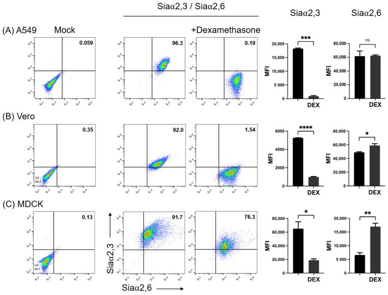
N-acetylneuraminic acid linked to galactose by α2,6 and α2,3 linkages (Siaα2,6 and Siaα2,3) is expressed on glycoconjugates of animal tissues, where it performs multiple biological functions. In addition, these types of sialic acid residues are the main targets for the binding and entry of influenza viruses. Here we used fluorochrome-conjugated Sambuccus nigra, Maackia amurensis, and peanut lectins for the simultaneous detection of Siaα2,3 and Siaα2,6 and galactosyl residues by two-color flow cytometry on A549 cells, a human pneumocyte cell line used for in vitro studies of the infection by influenza viruses, as well as on Vero and MDCK cell lines. The dexamethasone (DEX) glucocorticoid (GC), a widely used anti-inflammatory compound, completely abrogated the expression of Siaα2,3 in A549 cells and decreased its expression in Vero and MDCK cells; in contrast, the expression of Siaα2,6 was increased in the three cell lines. These observations indicate that DEX can be used for the study of the mechanism of sialylation of cell membrane molecules. Importantly, DEX may change the tropism of avian and human/pig influenza viruses and other infectious agents to animal and human epithelial cells.

Keywords: A549; MDCK; Vero; dexamethasone; influenza receptors; sialic acid; sialylation; zoonosis.


Conflict of interest statement

The authors declare that there are no conflict of interest.

Figures

Figure 1
Figure 1
Two-color flow cytometry analysis of the expression of (A) Siaα2,3 and Galβ1-3GalNAc and (B) Siaα2,6 and Galβ1-3GalNAc on A549 cells by means of binding of SNA, MAAII, and PNA lectins. The effect of treatment of the cell surface with neuraminidase, blocking of PNA binding with galactose, and lectin absorption with fetuin is shown. The percentage of positive cells in the upper right quadrants is shown. The mean fluorescence intensity of triplicates in a representative experiment is shown on the right. MFI, mean fluorescence intensity; Gal, galactose; Neu, neuraminidase; Fet, fetuin. Significant differences are marked with asterisks (**** p < 0.0001, one-way ANOVA).
Figure 2
Figure 2
Effect of neuraminidase on the expression of Siaα2,3 and Siaα2,6 on (A) A549, (B) Vero, and (C) MDCK cells. The effect of treatment of the cell surface with neuraminidase and lectin absorption with fetuin is shown. The mean fluorescence intensity of triplicates in a representative experiment is shown on the right. FMI, mean fluorescence intensity; Neu, neuraminidase; Fet, fetuin. Significant differences are marked with asterisks (p < 0.0001, one-way ANOVA).
Figure 3
Figure 3
Effect of treatment of cells with dexamethasone (DEX)on the expression of Siaα2,3 and Siaα2,6 on (A) A549, (B) Vero, and (C) MDCK cells. FMI, mean fluorescence intensity; DEX, dexamethasone; ns, non-significative difference. Significant differences are marked with asterisks (*, p < 0.05, **, p < 0.01, ***, p < 0.001, ****, p < 0.0001; Student’s t-test).
Figure 4
Figure 4
Output of the search in the SugarBind Database showing associations between pathogens and glycans. The query NeuAc(α2-6)Gal(β1-4)GlcNAc, the trisaccharide recognized by SNA, returned all pathogens matching these criteria. Influenza A H1, H3, H7, and H9 and Toxoplasma gondii recognize this glycan.

References

    1. Alvarez G., Lascurain R., Perez A., Degand P., Montano L.F., Martinez-Cairo S., Zenteno E. Relevance of sialoglycoconjugates in murine thymocytes during maturation and selection in the thymus. Immunol. Investig. 1999;28:9–18. doi: 10.3109/08820139909022719. - DOI - PubMed
    1. Woronowicz A., Amith S.R., De Vusser K., Laroy W., Contreras R., Basta S., Szewczuk M.R. Dependence of neurotrophic factor activation of Trk tyrosine kinase receptors on cellular sialidase. Glycobiology. 2007;17:10–24. doi: 10.1093/glycob/cwl049. - DOI - PubMed
    1. Kang J., Park H.M., Kim Y.W., Kim Y.H., Varghese S., Seok H.K., Kim Y.G., Kim S.H. Control of mesenchymal stem cell phenotype and differentiation depending on cell adhesion mechanism. Eur. Cell Mater. 2014;28:387–403. doi: 10.22203/eCM.v028a27. - DOI - PubMed
    1. Nightingale T.D., Frayne M.E., Clasper S., Banerji S., Jackson D.G. A mechanism of sialylation functionally silences the hyaluronan receptor LYVE-1 in lymphatic endothelium. J. Biol. Chem. 2009;284:3935–3945. doi: 10.1074/jbc.M805105200. - DOI - PubMed
    1. Takeuchi T., Sugimoto A., Imazato N., Tamura M., Nakatani S., Kobata K., Arata Y. Glucosamine Suppresses Osteoclast Differentiation through the Modulation of Glycosylation Including O-GlcNAcylation. Biol. Pharm. Bull. 2017;40:352–356. doi: 10.1248/bpb.b16-00877. - DOI - PubMed

LinkOut - more resources